-
PDF
- Split View
-
Views
-
Cite
Cite
Madhankumar Kuppuswamy, Philemon Gukop, George Sutherland, Chandrasekaran Venkatachalam, Kawasaki disease presenting as cardiac tamponade with ruptured giant aneurysm of the right coronary artery, Interactive CardioVascular and Thoracic Surgery, Volume 10, Issue 2, February 2010, Pages 317–318, https://doi.org/10.1510/icvts.2009.215731
Close - Share Icon Share
Abstract
We report a case of a 22-year-old man with Kawasaki disease presenting with features of cardiac tamponade following rupture of giant aneurysm of his right coronary artery. He underwent an emergency operation. Aneurysmal sac was of size 4×4 cm. The entry point of the aneurysm was sutured. Right coronary artery was grafted with left radial artery. He had an uneventful recovery in the postoperative period.
1. Case report
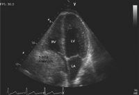
A 22-year-old gentleman presented with a history of dull aching chest pain of 6 h duration. He was diagnosed to have Kawasaki disease at the age of five years. He was known to have patent ductus arteriosus with aneurysm of the right coronary artery and was being followed-up in a different hospital. On examination his heart rate was 96 beats per min and blood pressure was 96/70 mmHg. His ECG showed Q wave with T wave inversion in the inferior leads. It also showed features of right bundle branch block. His troponin was negative. Echocardiography showed a mass over the right heart of size 4×4 cm with trivial pericardial effusion (Fig. 1 ). CT-scan showed a large mass of size 8×7 cm compressing the right atrium with calcified left coronary artery aneurysm. As the CT-scan was being performed his hemodynamics deteriorated with increasing size of the mass. Hence, he was taken for emergency surgery before completing the CT-scan.
Echocardiography showing thrombus in the right coronary artery aneurysm (T-RCA aneurysm) compressing right atrium.
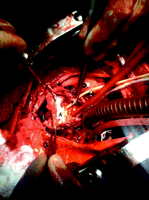
During surgery his left femoral artery was cannulated. Then the chest was opened by median sternotomy. Approximately 750 ml of blood with blood clots were evacuated from the pericardial cavity. Bleeding was initially controlled with the finger. Ruptured giant right coronary artery aneurysmal sac was identified. The aneurysmal sac was just adjacent to the right coronary artery ostium. Entry point of the right coronary artery aneurysm was identified and the bleeding was then controlled by introducing Foley's catheter into the ascending aorta through the entry point of the aneurysmal sac (Fig. 2 ). Two-stage single venous cannula was inserted to the right atrial appendage and the patient was connected to cardiopulmonary bypass. Patient's temperature was maintained at 34 °C throughout the procedure.
Intra-operative picture showing Foley's catheter (F) in the right coronary artery ostia and the right coronary artery aneurysmal sac (A).
The entry point of the aneurysmal sac was at the origin of right coronary artery (RCA) which was closed with two Teflon pledgetted sutures. The aneurysmal sac was measuring 4×4 cm. Distal opening of the aneurysmal sac was also identified and sutured. Patent ductus arteriosus was identified looped and ligated. In the meantime left radial artery was harvested. Distal right coronary artery was opened and endarterecomy was done (specimen sent for histopathological examination) as the posterior descending artery was of small calibre and not graftable. Distal anastomosis of the left radial artery to right coronary artery was done with heart in ventricular fibrillation. Proximal anastomosis completed. Calcified aneurysm of size 1.5 cm was noted in the proximal left anterior descending artery. There was no evidence of ischemia in the left anterior descending artery territory. Hence, left anterior descending artery was not grafted. The patient came off cardiopulmonary bypass without difficulty. He had an uneventful recovery in the postoperative period and was discharged on the fifth postoperative day. Postoperative echocardiogram done before discharge showed moderately impaired function of the right ventricle. No pericardial collection was noted.
2. Discussion
Kawasaki disease or mucocutaneous lymph node syndrome is a systemic disease with generalised vasculitis predominantly affecting infants and young children [1]. It affects primarily medium sized muscular arteries [2]. Damage caused by the disease in the pediatric age group can progress and present in the adolescent or adult age group. The involved arteries develop aneurysmal formation, thrombotic occlusion and premature atherosclerosis leading to ischemic heart disease [3]. Kato et al. showed the incidence of coronary aneurysm in acute Kawasaki disease was 25%, 55% of which showed regression. Giant coronary artery aneurysm was noted in 4.4% of the patients. During follow-up, ischemic heart disease developed in 4.7% and myocardial infarction in 1.9%. Death occurred in 0.8%. Death due to rupture of coronary artery aneurysm is extremely rare [3].
In untreated group, incidence of coronary artery aneurysm is 20% compared to 4–8% in the group treated with IV gamma immunoglobulin [4]. Hiroyuki et al. graded coronary artery aneurysm from grade 0 to grade 3, grade 3 being giant aneurysms with maximal diameter 8 mm. These grade 3 aneurysms are less likely to reduce in size and will often thrombose or rupture. Giant coronary artery aneurysms are very rare presentation [5].
In the case presented only the femoral artery was cannulated. Femoral vein was exposed but not cannulated. As the mass was compressing almost the entire right atrium, positioning the femoral cannula in the right atrium would have been difficult. Possibility of the femoral cannula disturbing the mass in the right atrium was also considered. As the clots were being removed from the pericardial cavity, the bleeding point was noted. It was controlled with the finger initially and the structures were identified. Then the patient was connected to cardiopulmonary bypass without significant hemodynamic deterioration. Fibrillatory arrest period was restricted to the distal anastomosis of the left radial artery to right coronary artery. All the other procedures were done without arresting the heart.
Calcification, fissuring, deposits of protein-like material and hyaline degeneration of the intima of the involved arteries in the Kawasaki disease were reported [6]. Histopathology of the endarterectomy specimen in the case reported showed marked fibrous scarring and calcification within the muscular wall. No active vasculitis or granulomas.
Giant coronary artery aneurysms which show increased rate of growth in size should be managed surgically in order to avoid acute complication like rupture as it happened in the case presented. Also, planning the surgery according to the need of the emergency situation like avoiding cardioplegic arrest in the case reported is to be considered for better outcome of these patients.


