Published online Aug 15, 2004. doi: 10.3748/wjg.v10.i16.2459
Revised: March 19, 2004
Accepted: April 9, 2004
Published online: August 15, 2004
AIM: Late unset of dysphagia due to vascular abnormalities is a rare condition. We aimed to present a case of right subclavian artery abnormalities caused dysphagia in the elderly.
METHODS: A 68-year-old female was admitted with dysphagia seven months ago. Upper endoscopic procedures and routine examinations could not demonstrate any etiology. Multislice computed thorax tomography was performed for probable extra- esophagial lesions.
RESULTS: Multislice computed thorax tomography showed right subclavian artery abnormality and esophagial compression with this aberrant artery.
CONCLUSION: Causes of dysphagia in the elderly are commonly malignancies, strictures and/or motility disorders. If routine examinations and endoscopic procedures fail to show any etiology, rare vascular abnormalities can be considered in such patients. Multislice computed tomography is a usefull choice in such conditions.
- Citation: Kantarceken B, Bulbuloglu E, Yuksel M, Cetinkaya A. Dysphagia lusorium in elderly: A case report. World J Gastroenterol 2004; 10(16): 2459-2460
- URL: https://www.wjgnet.com/1007-9327/full/v10/i16/2459.htm
- DOI: https://dx.doi.org/10.3748/wjg.v10.i16.2459
Dysphagia Lusoria was first described by Bayford in 1794 in a 62 year-old woman[1]. Postmortem examination of this case showed the abnormal origination of right subclavian artery from aortic arch and compression on the esophagus. Abnormalities of the thoracic aorta and great vessels are not uncommon and can result in esophageal compression and dysphagia. The most common congenital abnormality of the aorta is an isolated aberrant right subclavian artery[2]. Usually this abnormality does not lead to symptoms. However, sometimes dysphagia (dysphagia lusoria) develops. Mass effect on the esophagus can cause dysphagia. A right aortic arch with an aberrant left subclavian artery is less common but may also result in esophageal compression[3]. A pulmonary sliding occurs when an aberrant left pulmonary artery arises from the right pulmonary artery and passes between trachea and esophagus. Compression on both trachea and esophagus can occur. This abnormality can also be reliably detected with contrast-enhanced CT. We aimed to present a 68-year-old woman who had late onset dysphagia due to such a rare condition.
A 68-year-old female was admitted to our hospital with dysphagia nearly for seven months. Dysphagia was occuring both solid and liquids. There were no clear symptoms except dysphagia, such as loss of weight, fever, sweating at night, diarrhea, hematemesis, melena or hematochesia. She complained about odinophagia, bloating, regurgitation and epigastric pain especially after analgesic using. She also had chest pain radiating to the left arm with effort. She had a history of operation for discal hernia, ischemic heart disease and diabetes mellitus. She was using anti-ischemic drugs, analgesics (Including aspirin), beta blockers and diuretics.
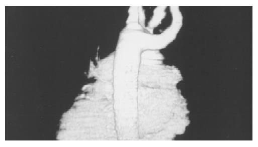
In physical examination her general condition was good, thyroid was palpeable. She had mild epigastric pain with palpation and no other signs. Hgb was136 g/L, WBC (White blood cell count) was 77000 μL, PLT was 290000 μL in laboratory tests. Erythrocyte sedimentation rate was 10 /h, SGOT was 57 U/L, SGPT was 84 U/L, ALP was 245 U/L. Other biochemical parameters were normal (BUN, creatinine, glucose, etc). Markers for hepatitis A,B,C were negative. Thyroid function tests were normal. She had esophagus graphy with radiopaque two months ago showing slight compression on esophagus in lower levels. Esophagogastroduodenoscopy (EGD) 5 mo ago showed minimal hiatus hernia, reflux esophagitis and antral gastritis. Some drugs were given to the patient for these findings but her dysphagia symptoms did not relieve. EGD 3 mo after, only showed antral gastritis. Other drugs (PPIs, antiacids) were given also, but dysphagia of the patient did not relieve. After admission to our clinic, the major complaint of the patient was persistent dysphagia despite the treatments. Thorax CT was performed (Multislice spiral CT) to exclude thoracal lesion-dysphagia, it showed right subclavian artery abnormality and esophagal compression on this aberrant artery (Figure 1A, B). Multislice computed thorax tomography (MCT) showed the right subclavian artery originating from the posterior wall of the aortic arch as its last branch distal to the origin of the left subclavian artery and it passed obliquely between esophagus and vertebral column and then coursed upwards on the right side.
Dysphagia is a common problem that lowers quality of life for the elderly and a symptom that may originate from many different causes. Esophageal dysphagia could be caused by esophageal carcinoma, esophageal stricture and webs, achalasia, diffuse esophageal spasm and scleroderma, caustic esophagitis and infectious esophagitis[4]. The other rare cause of dysphagia in the elderly is vascular compression on the esophagus (Dysphagia lusoria). Based on autopsy findings, the lusorian artery had a prevalence of 0.7% in the general population[5]. Recently Fockens et al[6] found a prevalence of 0.4% in 1629 patients who underwent endoscopy for various reasons. Dysphagia lusoria might be seen in young adults[7], and in middle and/or elder ages as in our case[8]. Why it gives symptoms in elderly is not clear. Some theories have been suggested, such as increased rigidity of the esophagus itself or the vessel wall, aneurysm formation, especially in the presence of a Kommerell’s diverticulum[9], elongation of the aorta, and the combination of an aberrant artery and a truncus bicaroticus[10]. Interestingly in our patient, she had a history of dysphagia for 7 mo. She had not any lesion except minimal esophagitis in EGD. Reflux esophagitis can explain dysphagia sometimes. After the treatment of esophagitis with proton pump inhibitors (PPI), our patient’s dysphagia symptom did not relieve. The late onset of dysphagia in our patient could be explained by the changes in vertebral column. Retrosternal goitre which may be responsable for dysphagia was not found in CT. Radiopaque graphy of the esophagus could be used to show the compression of aberrant artery on esophagus, but CT scanning and/or angiography can usually confirm the diagnosis of dysphagia lusoria. Aberrant artery could be shown with multislice CT as three dimension angiographic images were non-invasive without the need of invasive angiography[11]. Here, we also presented the multislice computed tomography angiography images of the patient with a symptomatic aberrant retro-esophagial subclavian artery compression (Figure 2).
We showed the aberrant right subclavian artery abnormality and compression on the esophagus clearly. That is why we did not repeat the radiopaque graphy of the esophagus. Coronary angiography was planned but the patient refused this procedure. So the patient was referred to a cardiovascular surgeon.
In conclusion, dysphagia in an elder patient can be caused by a rare abnormality of the subclavian artery insertion. Multislice CT can reveal this abnormality.
Edited by Wang XL Proofread by Xu FM
| 1. | Bayford D. An account of singular case of obstructed deglutition. Memoires Med Soc London 1794; 2: 275-286. . [Cited in This Article: ] |
| 2. | McLoughlin MJ, Weisbrod G, Wise DJ, Yeung HP. Computed tomography in congenital anomalies of the aortic arch and great vessels. Radiology. 1981;138:399-403. [PubMed] [DOI] [Cited in This Article: ] [Cited by in Crossref: 46] [Cited by in F6Publishing: 47] [Article Influence: 1.1] [Reference Citation Analysis (0)] |
| 3. | Jaffe RB. Radiographic manifestations of congenital anomalies of the aortic arch. Radiol Clin North Am. 1991;29:319-334. [PubMed] [Cited in This Article: ] |
| 4. | Barloon TJ, Bergus GR, Lu CC. Diagnostic imaging in the evaluation of dysphagia. Am Fam Physician. 1996;53:535-546. [PubMed] [Cited in This Article: ] |
| 5. | Molz G, Burri B. Aberrant subclavian artery (arteria lusoria): sex differences in the prevalence of various forms of the malformation. Evaluation of 1378 observations. Virchows Arch A Pathol Anat Histol. 1978;380:303-315. [PubMed] [Cited in This Article: ] |
| 6. | Fockens P, Kisman K, Tytgat GNJ. Endosonographic imaging of an aberrant right subclavian (lusorian) artery. Gastrointest Endosc. 1996;43:419. [DOI] [Cited in This Article: ] [Cited by in Crossref: 4] [Cited by in F6Publishing: 6] [Article Influence: 0.2] [Reference Citation Analysis (0)] |
| 7. | Ballotta E, Bardini R, Bottio T. Aberrant right subclavian artery. An original median cervical approach. J Cardiovasc Surg (Torino). 1996;37:571-573. [PubMed] [Cited in This Article: ] |
| 8. | Morris CD, Kanter KR, Miller JI. Late-onset dysphagia lusoria. Ann Thorac Surg. 2001;71:710-712. [PubMed] [DOI] [Cited in This Article: ] [Cited by in Crossref: 10] [Cited by in F6Publishing: 13] [Article Influence: 0.6] [Reference Citation Analysis (0)] |
| 9. | Brown DL, Chapman WC, Edwards WH, Coltharp WH, Stoney WS. Dysphagia lusoria: aberrant right subclavian artery with a Kommerell's diverticulum. Am Surg. 1993;59:582-586. [PubMed] [Cited in This Article: ] |
| 10. | Janssen M, Baggen MG, Veen HF, Smout AJ, Bekkers JA, Jonkman JG, Ouwendijk RJ. Dysphagia lusoria: clinical aspects, manometric findings, diagnosis, and therapy. Am J Gastroenterol. 2000;95:1411-1416. [PubMed] [DOI] [Cited in This Article: ] [Cited by in Crossref: 97] [Cited by in F6Publishing: 105] [Article Influence: 4.4] [Reference Citation Analysis (0)] |
| 11. | Morgan-Hughes GJ, Owens PE, Roobottom CA. Aberrant right subclavian artery and dysphagia lusoria. N Engl J Med. 2002;347:1532. [PubMed] [Cited in This Article: ] |


