
Abstract
Benign and primary malignant breast tumors are quite common, but secondary tumors in the breast from metastatic malignancies are rare. Nevertheless, metastasis to the breast must be considered in any patient with a known primary malignant tumor history who presents with a breast lump. We report a case of a premenopausal woman diagnosed with a metastatic melanoma in her breast.
INTRODUCTION
Besides hosting many benign and primary malignant tumors, the breast is also a metastatic site for tumors from leukemia, lung cancer, and melanoma.1-4 Secondary tumors in the breast represent 1.3%-2.7% of all malignant mammary tumors.5 We report the case of a premenopausal woman who was diagnosed with a metastatic melanoma in her breast.
CASE REPORT
A 42-year-old premenopausal Caucasian woman was referred to our breast clinic with a 10-day history of a left breast lump. The patient had a history of a primary cutaneous melanoma (1.1 mm thick according to Breslow classification) removed from the right ankle 4 years before her presentation with the breast lump. Three years later, she had a single cutaneous recurrence in the medial side of the right thigh that was locally excised. No further treatment was required because there was no evidence of metastasis following the melanoma staging process. She was monitored clinically and by imaging (full-body computed tomography [CT] scan and magnetic resonance imaging of the brain). These images showed no evidence of melanoma recurrence up to 2 months prior to her presentation with the breast lump.
Examination found a hard lump in the upper outer quadrant of the left breast measuring 2 × 2 cm, with a few palpable lymph nodes in the left axilla. The overlying skin was normal. She also had a recurrent skin lesion on her back.
A mammogram showed an indeterminate mass in the left breast (M3), while a left breast ultrasound indicated that the mass was a highly suspicious lobulated (U4) lesion (Figure 1). From our experience, metastatic lesions in the breast have unusual and potentially confusing appearances on imaging. On mammograms they tend to be fairly well defined, and on ultrasound they can be lobulated. However, very few breast primary malignancies show these features.

Mammogram of the left breast showing a well-defined mass (M). Ultrasound scan of the left breast showing a multilobulated mass (U).
In addition, the patient's left axilla ultrasound showed an enlarged lymph node with a thick cortex but fatty hilum.
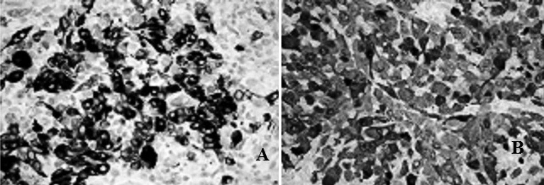
A fine needle aspiration (FNA) and biopsy of the left breast lump yielded C5 (malignant) cytology, and the core biopsy showed a poorly differentiated malignancy. Melanoma markers showed 100% positivity to the S-100 protein and patchy staining for Melan-A and HMB-45, as seen in Figure 2. FNA of the left axillary lymph node revealed the node to be C2 (benign).

Melanoma stains of the left breast lump core biopsy. A: 100% positivity to S-100 protein. B: Patchy staining for Melan-A and HMB-45.
A multidisciplinary team discussed the above triple assessment (clinical, radiologic, and histopathologic) and diagnosed breast metastasis from a melanoma. After we found no further evidence of disease spread, the patient underwent a local excision of the breast lump and the back lesion. The histopathology report for both lesions confirmed features of metastatic melanoma.
A recurrent skin lesion developed on her left buttock a few months later; the patient also experienced brain metastasis. Both were surgically removed. The most recent CT scan showed enlarged hilar lymph nodes. Currently, she is participating in an oncology clinical trial involving chemotherapy.
DISCUSSION
Melanoma is the most rapidly increasing cancer in Caucasians, and 20% of patients diagnosed with melanoma will develop metastasis via hematogenic or lymphatic routes. Melanoma can spread to lymph nodes, secondary sites in the skin, and distant organs such as the breast.1,6-8
Melanoma in the breast could be primary in the breast skin, primary in the breast tissue, metastasis in the breast, or in-transit metastases to breast tissue and breast skin.1 The most common site for primary melanoma in premenopausal women is the lower extremities; in comparison, the most common primary sites for melanoma in premenopausal women with melanoma metastasis in the breast are the trunk and upper limb.3,7
Metastasis is more common in the outer half of the breast because of good vascularity and the presence of more glandular tissue.9,10 A review of metastasis to the breast from nonmammary tumors by Toombs and Kalisher found that 50% of metastases were located in the upper outer quadrant of the breast.11,12 The outer breast is also a common site for the development of primary breast cancer (66% of patients). Therefore, the site of a lump does not help differentiate primary and secondary malignancies.13,14
An autopsy series of 1,000 breast cancer patients found metastatic melanoma in the breast in 5% of the cases.15 Secondary breast lesions could be the first manifestation of melanoma, with metastasis to the breast in 40% of the affected patients.3
In metastatic melanoma, a full-body CT scan is recommended. Positron emission tomography is unlikely to be clinically relevant in established metastasis, but it may be useful in excluding disease that might make surgery inappropriate.16
Elective lymph node dissection has no place in the management of primary melanoma unless it is unavoidable because the lesion lies over the lymph node. In clinically negative lymph nodes, sentinal lymph node biopsy might be used as a staging procedure; however, it has no proven therapeutic value and carries a 5% morbidity risk. FNA of the lymph nodes (with or without core biopsy) is recommended for clinically or radiologically suspicious lymph nodes. Open biopsy is sometimes performed for suspicious lymph nodes (even with negative FNA). In such cases, the incision must allow subsequent complete formal block dissection of the regional nodes without compromise.16
The overall survival for patients with metastatic melanoma ranges from 4.7 to 11 months.17 Treatment options include close observation, surgical resection of isolated metastases (skin, brain, gut), therapy with dacarbazine for palliation, and participation in clinical trials.16,17 Radiotherapy may have a palliative role in the treatment of metastases.
A recently published randomized controlled trial has shown that the BRAF kinase inhibitor vemurafenib improved rates of overall and progression-free survival in patients with previously untreated melanoma with the BRAF V600E mutation.18
CONCLUSIONS
Metastasis to the breast must be considered in any patient with a known primary malignant tumor history who presents with a breast lump. Careful triple assessment and multidisciplinary decisionmaking are vital in developing the management plan.
This article meets the Accreditation Council for Graduate Medical Education and the American Board of Medical Specialties Maintenance of Certification competencies for Patient Care and Medical Knowledge.
Footnotes
The authors have no financial or proprietary interest in the subject matter of this article.
- Academic Division of Ochsner Clinic Foundation


