
Main menu

  • Home
  • Content
    • Current
    • Ahead of print
    • Archive
  • Info for
    • Authors
    • Reviewers
  • About Us
    • About the Ochsner Journal
    • Editorial Board
  • More
    • Alerts
    • Feedback
  • Other Publications
    • Ochsner Journal Blog

User menu

  • My alerts
  • Log in

Search

  • Advanced search
Ochsner Journal
  • Other Publications
    • Ochsner Journal Blog
  • My alerts
  • Log in
Ochsner Journal

Advanced Search

  • Home
  • Content
    • Current
    • Ahead of print
    • Archive
  • Info for
    • Authors
    • Reviewers
  • About Us
    • About the Ochsner Journal
    • Editorial Board
  • More
    • Alerts
    • Feedback
Case ReportCase Reports and Clinical Observations

Pyoderma Gangrenosum: A Rare Cause of Breast Ulceration

Georgina Duke, Ahmad Al Samaraee, Akhtar Husain, Simon Meggitt and Tarannum Fasih
Ochsner Journal June 2012, 12 (2) 155-158;
Georgina Duke
*South Tees Hospitals National Health Service Foundation Trust, Middlesbrough, UK
  • Find this author on Google Scholar
  • Find this author on PubMed
  • Search for this author on this site
Ahmad Al Samaraee
†South Tyneside District Hospital, South Shields, UK
  • Find this author on Google Scholar
  • Find this author on PubMed
  • Search for this author on this site
Akhtar Husain
‡Newcastle Hospitals National Health Service Foundation Trust, Newcastle, UK
  • Find this author on Google Scholar
  • Find this author on PubMed
  • Search for this author on this site
Simon Meggitt
‡Newcastle Hospitals National Health Service Foundation Trust, Newcastle, UK
  • Find this author on Google Scholar
  • Find this author on PubMed
  • Search for this author on this site
Tarannum Fasih
§Queen Elizabeth Hospital, Gateshead, UK
  • Find this author on Google Scholar
  • Find this author on PubMed
  • Search for this author on this site
  • Article
  • Figures & Data
  • References
  • Info & Metrics
  • PDF
Loading

Abstract

Breast ulceration is an alarming sign for clinicians and places a significant physical and psychological burden on the patient. We report a rare presentation of pyoderma gangrenosum of the breast in a patient known to have ulcerative colitis but no active underlying disease process and no history of breast tissue trauma. This case report with literature review highlights the importance of considering pyoderma gangrenosum as a differential diagnosis in breast ulcers.

Keywords
  • Breast ulcer
  • necrotizing skin disease
  • pyoderma gangrenosum

INTRODUCTION

Although breast cancer would be the most likely differential clinical diagnosis in any patient with breast ulceration, other rare causes should be considered. We present a case report about a postmenopausal woman with breast ulceration caused by pyoderma gangrenosum (PG).

CASE REPORT

A 54-year-old postmenopausal white woman presented with a very painful left breast ulcer of 2 weeks' duration. The ulcer started as a small boil with yellow discharge and then increased gradually in size. She had no history of recent gastrointestinal or other systemic symptoms and had not previously undergone any surgery or biopsy of her left breast.

The patient had been diagnosed with ulcerative colitis (UC) 19 years earlier. Her last colonoscopy was a few months before her presentation; the results did not show features of active colitis. She was on long-term sulphasalazine (3 g/day) for her colitis. She stopped smoking at the age of 35 and had no family history of breast disease.

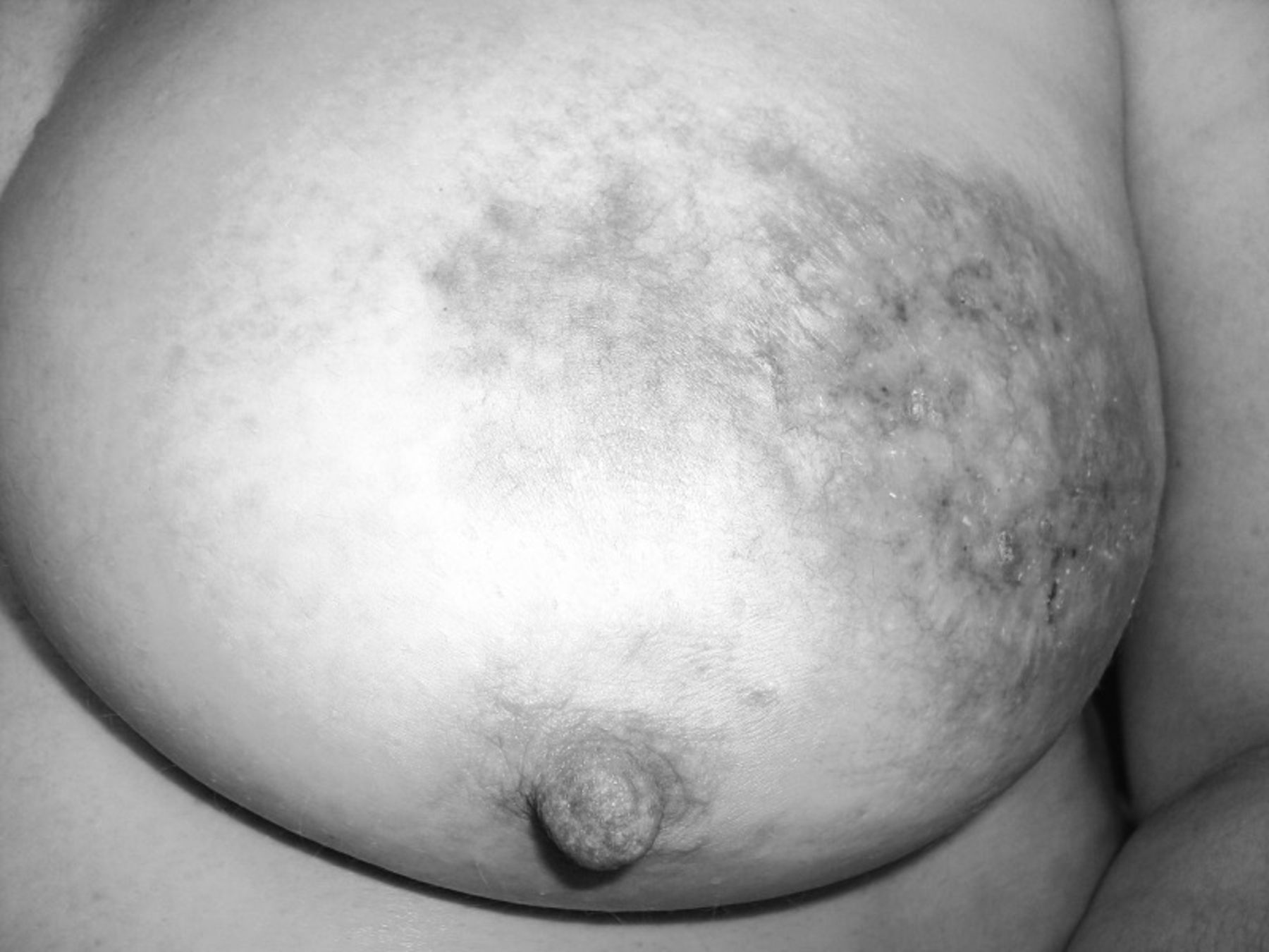
Clinical examination revealed an approximately 5 × 3 cm superficial ulcer located mainly in the upper central part of the left breast. The ulcer was very tender and surrounded by erythema and induration with yellow discharge (Figure 1). Nothing else was found during examination of both breasts and axillae. Abdominal examination was unremarkable.

Figure 1.
  • Download figure
  • Open in new tab
  • Download powerpoint
Figure 1.

Pretreatment: left breast pyoderma gangrenosum.

Routine blood tests showed normal liver and renal function. The hemoglobin level was 11.3 gm/dL with a normal mean cell volume of 88.9 fL. The white cell and platelet counts were within the normal range. The erythrocyte sedimentation rate (ESR) was slightly elevated at 24 mm/hr, and the autoantibody screening test was unremarkable. The culture and sensitivity test of the ulcer microbiology swab was negative. An ultrasound scan of both breasts was normal. We did not perform mammography because we believed it would be too painful.

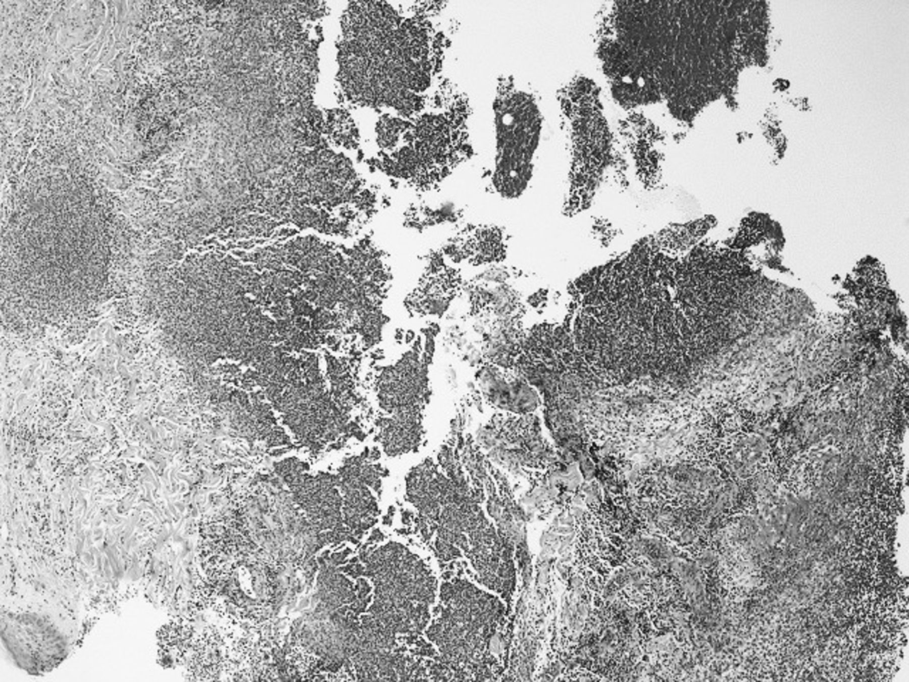
A punch biopsy of the ulcer showed occasional superficial subcorneal pustules. The epidermis was acanthotic and spongiotic with inflammatory exocytosis. The dermis contained a dense, diffuse neutrophilic infiltrate. We found no evidence of malignancy. The overall features were those of neutrophilic inflammation with abscess formation. The histologic features were not specific but were compatible with the clinical diagnosis of PG (Figures 2 and 3).

Figure 2.
  • Download figure
  • Open in new tab
  • Download powerpoint
Figure 2.

Ulcer edge with evidence of active inflammation and abscess formation in the dermis composed of polymorphs (hematoxylin-eosin stain, magnification ×25).

Figure 3.
  • Download figure
  • Open in new tab
  • Download powerpoint
Figure 3.

High-power view of the abscess shows large numbers of polymorphs and granulation tissue reaction (hematoxylin-eosin stain, magnification ×40).

The patient was treated with 50 mg prednisolone daily and topical corticosteroids (0.1% betamethasone cream) for the first 3 months; the oral dose has since been reduced. She is currently on 10 mg prednisolone daily. This treatment has resulted in a significant improvement of the ulcer (Figure 4). Her most recent ESR was 18 mm/hr. She is still being seen for regular follow-up, and the oral steroid dose is monitored according to the clinical response.

Figure 4.
  • Download figure
  • Open in new tab
  • Download powerpoint
Figure 4.

Posttreatment: left breast pyoderma gangrenosum.

DISCUSSION

PG is a rare cutaneous necrotising skin disease characterised by a chronic neutrophilic inflammatory reaction and resulting in erythematous papules with blue-purple-red margins, most often presenting as painful ulceration.1,2

Brocq1 first described this idiopathic process in 1916 as occurring in all age groups but most commonly in patients in their 30s through 50s with a similar sex distribution. The overall incidence is unknown because of the rarity of the condition, but the literature estimates it to be 3:1,000,000 annually in the United States.3

The pathogenesis of the process is unknown, but suggested causes include immune-complex mediated neutrophilic vascular reactions. The diagnosis is predominantly clinical; histopathologic findings and laboratory tests serve mostly to rule out mimicking lesions. The presence or absence of vasculitis is a controversial feature but is not thought to be a primary event even if present. The earliest lesions show follicular and perifollicular inflammation with neutrophilic abscesses in the dermis. These lesions can later lead to dermal necrosis with ulceration and undermining inflammation at the ulcer edge.1,2,4-7

The most common anatomical site for PG in patients with inflammatory bowel disease (IBD) is the lower limbs, where it has been reported in 75%-80% of IBD patients.1,8-10 PG was originally thought to be pathognomonic of IBD, affecting UC patients more than those with Crohn disease.2,11-14 Although this connection is commonly true, other conditions have been associated with PG, such as rheumatoid arthritis and hematologic disorders.1,15-20 The prevalence rate of associated systemic disease ranges widely in the literature (50%-78%).1,3,21-24

Reported therapies for PG have often been focused on treatment of the underlying condition; therefore, most information available relates to systemic therapies,2,25,26 probably because many PG lesions mimic the activity of the underlying disease.27 However, our patient had no symptoms of active UC throughout the clinical course of the PG lesion.

First-line therapies include immunosuppressants such as corticosteroids (prednisolone/methylprednisolone) and cyclosporin.2,13,28-30 Other therapies include immunomodulators, biologics, and antileprosy drugs. These treatments have been used predominantly on steroid-refractory PG lesions associated with IBD.1,30 As an adjunct to other treatment, systemic antibiotics may have a role in PG remission.1,31-33 Patients similar to ours, in whom the underlying associated disease is quiescent, can respond variably to standard therapy; PG can even recur after definitive surgery for the underlying disease, for example, colectomy.26,34-36

Angiò et al26 described a case of PG of the sinus mammarum in a patient with UC that emphasized the resistance of the cutaneous ulcer to conventional systemic medical therapy although the treatment was efficacious regarding the colonic process. They used the histopathologic finding of vasculitis at the edge of the lesions to justify the use of antithrombotic topical injections as an adjunct to standard treatment. This isolated case report suggested that perilesional injections of calcic heparin were successful in inducing remission.26

The role of topical therapies is not well evaluated in the literature.1,26 One retrospective analysis of 20 patients showed that topical steroids have little role in healing.1,37 Conversely, other authors have demonstrated improvement of the lesions with intralesional steroids (triamcinolone acetonide).38

Currently, the Study of Treatments for Pyoderma Gangrenosum Patients (STOP GAP) Trial is taking place in approximately 50 hospitals throughout the United Kingdom and Ireland, with the aim to investigate the best way to treat PG lesions. A total of 140 patients will be randomized to have either prednisolone or cyclosporin.39 Patients who initially require topical therapy will enter a parallel observational study. If their PG lesions fail to respond to topical therapy, the participants could be enrolled in the randomized trial of systemic treatments.40

Reported cases of surgical debridement and split skin grafts for PG lesions generally have poor outcomes. Perhaps these poor outcomes result from the pathergy phenomenon, a key feature in the disease process in which any traumatized skin (debridement sites or skin graft donor sites) develops additional necrosis and ulceration.41,42 Therefore, surgical intervention is not recommended in standard practice.

A review of published works disclosed 43 reported cases of PG of the breast, of which 30 cases (approximately 70%) reported that PG lesions were secondary to trauma of the breast tissue such as surgical intervention, reconstruction of the breast, biopsy, and intramuscular injection.1,2,20,26,43-48 Again, the pathergy phenomenon may explain the high percentage of PG lesions after breast trauma.

CONCLUSIONS

Our patient had a rare presentation of PG of the breast, in which she had associated UC but no active underlying disease process and no history of breast tissue trauma. This case highlights the importance of considering PG as one of the differential diagnoses of breast ulcers. Careful specialist clinical assessment could establish an early diagnosis and formulate an effective management plan. Long-term dermatology follow-up and contribution to clinical trials could provide a better understanding of the disease and its response to various treatment options.

This article meets the Accreditation Council for Graduate Medical Education and the American Board of Medical Specialties Maintenance of Certification competencies for Patient Care and Medical Knowledge.

Footnotes

  • The authors have no financial or proprietary interest in the subject matter of this article.

  • Academic Division of Ochsner Clinic Foundation

REFERENCES

  1. ↵
    1. Hasselmann DO,
    2. Bens G,
    3. Tilgen W,
    4. Reichrath J
    (7, 2007) Pyoderma gangrenosum: clinical presentation and outcome in 18 cases and review of the literature. J Dtsch Dermatol Ges 5(7):560–564, pmid:17610605.
    OpenUrlCrossRefPubMed
  2. ↵
    1. Tromm A,
    2. May D,
    3. Almus E,
    4. et al.
    (2, 2001) Cutaneous manifestations in inflammatory bowel disease. Z Gastroenterol 39(2):137–144, pmid:11253504.
    OpenUrlCrossRefPubMed
  3. ↵
    1. Powell FC,
    2. Schroeter AL,
    3. Su WP,
    4. Perry HO
    (7, 1984) Pyoderma gangrenosum and sarcoidosis. Arch Dermatol 120(7):959–960, pmid:6732265.
    OpenUrlCrossRefPubMed
  4. ↵
    1. Jorizzo JL,
    2. Solomon AR,
    3. Zanolli MD,
    4. Leshin B
    (12, 1988) Neutrophilic vascular reactions. J Am Acad Dermatol 19(6):983–1005, pmid:3060489.
    OpenUrlPubMed
    1. Newell LM,
    2. Malkinson FD
    (10, 1982) Commentary: pyoderma gangrenosum. Arch Dermatol 118(10):769–773.
    OpenUrlCrossRef
    1. Powell FC,
    2. Schroeter AL,
    3. Su WP,
    4. Perry HO
    (5, 1985) Pyoderma gangrenosum: a review of 86 patients. Q J Med 55(217):173–186, pmid:3889978.
    OpenUrlPubMed
  5. ↵
    1. Ackerman AB
    (1983;) Questions to the Editorial Board. Am J Dermatopath 5(4):409–410.
    OpenUrl
  6. ↵
    1. Levitt MD,
    2. Ritchie JK,
    3. Lennard-Jones JE,
    4. Phillips RK
    (6, 1991) Pyoderma gangrenosum in inflammatory bowel disease. Br J Surg 78(6):676–678, pmid:2070231.
    OpenUrlCrossRefPubMed
    1. von den Driesch P
    (12, 1997) Pyoderma gangrenosum: a report of 44 cases with follow-up. Br J Dermatol 137(6):1000–1005, pmid:9470924.
    OpenUrlCrossRefPubMed
  7. ↵
    1. Bennett ML,
    2. Jackson JM,
    3. Jorizzo JL,
    4. Fleischer AB Jr.,
    5. White WL,
    6. Callen JP
    (1, 2000) Pyoderma gangrenosum. A comparison of typical and atypical forms with an emphasis on time to remission. Case review of 86 patients from 2 institutions. Medicine (Baltimore) 79(1):37–46, pmid:10670408.
    OpenUrlCrossRefPubMed
  8. ↵
    1. Greenstein AJ,
    2. Janowitz HD,
    3. Sachar DB
    (9, 1976) The extra-intestinal complications of Crohn's disease and ulcerative colitis: a study of 700 patients. Medicine (Baltimore) 55(5):401–412, pmid:957999.
    OpenUrlCrossRefPubMed
    1. Kirsch B,
    2. Gerhardt H,
    3. Gladisch R,
    4. Heine M,
    5. Rohr G,
    6. Weiss J
    (1992;) Dermatosen bei chronisch entzündlichen Darmerkrankungen. Ergebnisse einer Untersuchung Von 119 Morbus-Crohn-/colitis Patienten. Akt Dermatol 18((1-2)):17–22.
    OpenUrl
  9. ↵
    1. Lamers CB
    (9, 1997) Treatment of extraintestinal complications of ulcerative colitis. Eur J Gastroenterol Hepatol 9(9):850–853, pmid:9355781.
    OpenUrlPubMed
  10. ↵
    1. Shanahan F
    1. McCord ML,
    2. Hall RP
    (1994) in Targan Sr in Inflammatory Bowel Disease: From Bench to Bedside, Cutaneous manifestations of inflammatory bowel disease, ed Shanahan F (Williams & Wilkins; Baltimore, MD), pp 682–694.
  11. ↵
    1. Bargen JA
    (10, 1929) Complications and sequelae of chronic ulcerative colitis. Ann Intern Med 3(4):335–352.
    OpenUrlCrossRef
    1. Crowson AN,
    2. Mihm MC Jr.,
    3. Magro C
    (2, 2003) Pyoderma gangrenosum: a review. J Cutan Pathol 30(2):97–107, pmid:12641787.
    OpenUrlCrossRefPubMed
    1. Tay CH
    (10, 1973) Letter: pyoderma gangrenosum and leukemia. Arch Dermatol 108(4):580–581, pmid:4517969.
    OpenUrlPubMed
    1. Jacobs P,
    2. Palmer S,
    3. Gordon-Smith EC
    (8, 1985) Pyoderma gangrenosum in myelodysplasia and acute leukaemia. Postgrad Med J 61(718):689–694, pmid:4034458.
    OpenUrlAbstract/FREE Full Text
    1. Struthers GR
    (4, 1979) Pyoderma gangrenosum, seronegative polyarthropathy and inflammatory bowel disease. J R Soc Med 72(4):284–286, pmid:552518.
    OpenUrlCrossRefPubMed
  12. ↵
    1. Labat JP,
    2. Simon H,
    3. Metges JP,
    4. Lucas B,
    5. Malhaire JP
    (6, 2000) [Pyoderma gangrenosum and breast cancer: a new case]. Ann Med Interne (Paris) 151(4):314–315, pmid:10922963, French.
    OpenUrlPubMed
  13. ↵
    1. Perry HO,
    2. Brunsting LA
    (3, 1957) Pyoderma gangrenosum; a clinical study of nineteen cases. AMA Arch Derm 75(3):380–386, pmid:13402210.
    OpenUrlPubMed
    1. Callen JP
    (2 21, 1998) Pyoderma gangrenosum. Lancet 351(9102):581–585, pmid:9492798.
    OpenUrlCrossRefPubMed
    1. Perry HO,
    2. Winkelmann RK
    (12, 1972) Bullous pyoderma gangrenosum and leukemia. Arch Dermatol 106(6):901–905, pmid:4508849.
    OpenUrlCrossRefPubMed
  14. ↵
    1. Prystowsky JH,
    2. Kahn SN,
    3. Lazarus GS
    (1, 1989) Present status of pyoderma gangrenosum. Review of 21 cases. Arch Dermatol 125(1):57–64, pmid:2642682.
    OpenUrlCrossRefPubMed
  15. ↵
    1. Thornton JR,
    2. Teague RH,
    3. Low-Beer TS,
    4. Read AE
    (3, 1980) Pyoderma gangrenosum and ulcerative colitis. Gut 21(3):247–248, pmid:7399325.
    OpenUrlAbstract/FREE Full Text
  16. ↵
    1. Angiò LG,
    2. Pirrone G,
    3. Rivoli G,
    4. et al.
    (Jun-Jul 2003) Pyoderma gangrenosum of the “sinus mammarum” in ulcerative colitis. G Chir 24((6-7)):247–254, pmid:14569923.
    OpenUrlPubMed
  17. ↵
    1. Lukash WM,
    2. Johnson RB
    1. Johnson WT,
    2. Norva WM
    (1975) in The Systemic Manifestations of Inflammatory Bowel Disease, Cutaneous manifestations of inflammatory bowel disease, eds Lukash WM, Johnson RB (Charles C. Thomas Publisher, Ltd, Springfield, IL), p 212.
  18. ↵
    1. Chow RK,
    2. Ho VC
    (6, 1996) Treatment of pyoderma gangrenosum. J Am Acad Dermatol 34(6):1047–1060, pmid:8647970.
    OpenUrlCrossRefPubMed
    1. Schwaegerle SM,
    2. Bergfeld WF,
    3. Senitzer D,
    4. Tidrick RT
    (3, 1988) Pyoderma gangrenosum: a review. J Am Acad Dermatol 18(3):559–568, pmid:3280623.
    OpenUrlCrossRefPubMed
  19. ↵
    1. Reichrath J,
    2. Bens G,
    3. Bonowitz A,
    4. Tilgen W
    (8, 2005) Treatment recommendations for pyoderma gangrenosum: an evidence-based review of the literature based on more than 350 patients. J Am Acad Dermatol 53(2):273–283, pmid:16021123.
    OpenUrlCrossRefPubMed
  20. ↵
    1. Wines N,
    2. Wines M,
    3. Ryman W
    (6 27, 2001) Understanding pyoderma gangrenosum: a review. MedGenMed 3(3):6, pmid:11549979.
    OpenUrlPubMed
    1. Powell FC,
    2. Su WP,
    3. Perry HO
    (3, 1996) Pyoderma gangrenosum: classification and management. J Am Acad Dermatol 34(3):395–409, 410–412, pmid:8609250, quiz.
    OpenUrlCrossRefPubMed
  21. ↵
    1. Davies MG,
    2. Piper S
    (3, 1981) Pyoderma gangrenosum: successful treatment with minocycline. Clin Exp Dermatol 6(2):219–223, pmid:7261471.
    OpenUrlCrossRefPubMed
  22. ↵
    1. Holmlund DE,
    2. Wählby L
    (1, 1987) Pyoderma gangrenosum after colectomy for inflammatory bowel disease. Case report. Acta Chir Scand 153(1):73–74, pmid:3577572.
    OpenUrlPubMed
    1. Kirsner JB,
    2. Shorter RG
    1. Mayer L,
    2. Janowitz HD
    (1988) in Inflammatory Bowel Disease, Extraintestinal manifestations of inflammatory bowel disease, eds Kirsner JB, Shorter RG (Philadelphia, PA), p 299.
  23. ↵
    1. Talansky AL,
    2. Meyers S,
    3. Greenstein AJ,
    4. Janowitz HD
    (6, 1983) Does intestinal resection heal the pyoderma gangrenosum of inflammatory bowel disease? J Clin Gastroenterol 5(3):207–210, pmid:6863878.
    OpenUrlCrossRefPubMed
  24. ↵
    1. Sheldon DG,
    2. Sawchuk LL,
    3. Kozarek RA,
    4. Thirlby RC
    (5, 2000) Twenty cases of peristomal pyoderma gangrenosum: diagnostic implications and management. Arch Surg 135(5):564–568, pmid:10807281, discussion 568-569.
    OpenUrlCrossRefPubMed
  25. ↵
    1. Goldstein F,
    2. Krain R,
    3. Thornton JJ
    (12, 1985) Intralesional steroid therapy of pyoderma gangrenosum. J Clin Gastroenterol 7(6):499–501, pmid:4086744.
    OpenUrlCrossRefPubMed
  26. ↵
    1. The Stop Gap Trial
    (2011) UK Dermatology Clinical Trials Network, July, http://www.stopgaptrial.co.uk/. Accessed 01 October 2011.
  27. ↵
    1. Mitchell E
    (2010;) RCT of treatments for Pyoderma gangrenosum: time to get involved. Wounds UK 6(4):27–32.
    OpenUrl
  28. ↵
    1. Kaddoura IL,
    2. Amm C
    (1, 2001) A rationale for adjuvant surgical intervention in pyoderma gangrenosum. Ann Plast Surg 46(1):23–28, pmid:11192029.
    OpenUrlCrossRefPubMed
  29. ↵
    1. Long CC,
    2. Jessop J,
    3. Young M,
    4. Holt PJ
    (7, 1992) Minimizing the risk of post-operative pyoderma gangrenosum. Br J Dermatol 127(1):45–48, pmid:1637694.
    OpenUrlPubMed
  30. ↵
    1. Mansur AT,
    2. Balaban D,
    3. Göktay F,
    4. Takmaz S
    (1, 2010) Pyoderma gangrenosum on the breast: a case presentation and review of the published work. J Dermatol 37(1):107–110, pmid:20175832.
    OpenUrlPubMed
    1. Duval A,
    2. Boissel N,
    3. Servant JM,
    4. Santini C,
    5. Petit A,
    6. Vignon-Pennamen MD
    (1, 2011) Pyoderma gangrenosum of the breast: a diagnosis not to be missed. J Plast Reconstr Aesthet Surg 64(1):e17–e20, pmid:20851068, Epub 2010 Sep 18.
    OpenUrlPubMed
    1. Goshtasby PH,
    2. Chami RG,
    3. Johnson RM
    (3, 2010) A novel approach to the management of pyoderma gangrenosum complicating reduction mammaplasty. Aesthet Surg J 30(2):186–193, pmid:20442095.
    OpenUrlCrossRefPubMed
    1. Maigre M,
    2. Bouachour G,
    3. Varache N,
    4. Alquier P,
    5. Verret JL
    (Nov-Dec 1991) [Pseudo-septicemic pyoderma gangrenosum and breast cancer. Apropos of a case caused by an intramuscular injection]. Rev Med Interne 12(6):452–454, pmid:1792438, French.
    OpenUrlPubMed
    1. Beurey J,
    2. Weber M,
    3. Delrous JL,
    4. Chaulieu Y
    (10, 1977) [Pyoderma gangrenosum: Clofazimine therapy]. Ann Dermatol Venereol 104(10):631–634, pmid:610518, French.
    OpenUrlPubMed
  31. ↵
    1. Caterson SA,
    2. Nyame T,
    3. Phung T,
    4. Lee B,
    5. Tobias AM
    (9, 2010) Pyoderma gangrenosum following bilateral deep inferior epigastric perforator flap breast reconstruction. J Reconstr Microsurg 26(7):475–479, pmid:20535668, Epub 2010 Jun 9.
    OpenUrlPubMed
PreviousNext
Back to top

In this issue

Ochsner Journal
Vol. 12, Issue 2
Jun 2012
  • Table of Contents
  • Index by author
Print
Download PDF
Email Article

Thank you for your interest in spreading the word on Ochsner Journal.

NOTE: We only request your email address so that the person you are recommending the page to knows that you wanted them to see it, and that it is not junk mail. We do not capture any email address.

Enter multiple addresses on separate lines or separate them with commas.
Pyoderma Gangrenosum: A Rare Cause of Breast Ulceration
(Your Name) has sent you a message from Ochsner Journal
(Your Name) thought you would like to see the Ochsner Journal web site.
CAPTCHA
This question is for testing whether or not you are a human visitor and to prevent automated spam submissions.
Citation Tools
Pyoderma Gangrenosum: A Rare Cause of Breast Ulceration
Georgina Duke, Ahmad Al Samaraee, Akhtar Husain, Simon Meggitt, Tarannum Fasih
Ochsner Journal Jun 2012, 12 (2) 155-158;

Citation Manager Formats

  • BibTeX
  • Bookends
  • EasyBib
  • EndNote (tagged)
  • EndNote 8 (xml)
  • Medlars
  • Mendeley
  • Papers
  • RefWorks Tagged
  • Ref Manager
  • RIS
  • Zotero
Share
Pyoderma Gangrenosum: A Rare Cause of Breast Ulceration
Georgina Duke, Ahmad Al Samaraee, Akhtar Husain, Simon Meggitt, Tarannum Fasih
Ochsner Journal Jun 2012, 12 (2) 155-158;
Reddit logo Twitter logo Facebook logo Mendeley logo
  • Tweet Widget
  • Facebook Like
  • Google Plus One

Jump to section

  • Article
    • Abstract
    • INTRODUCTION
    • CASE REPORT
    • DISCUSSION
    • CONCLUSIONS
    • Footnotes
    • REFERENCES
  • Figures & Data
  • Info & Metrics
  • References
  • PDF

Cited By...

  • Pyoderma gangrenosum of the breast 17 years after breast cancer treatment
  • Google Scholar

More in this TOC Section

  • Differential to Terminal Ileitis: Terminal Ileum Neuroendocrine Tumor Identified on Screening Colonoscopy
  • Pelvic Synovial Sarcoma Clinically Masquerading as an Ovarian Malignancy
  • Two-Stage Pediatric Ear Reconstruction Using Preserved Native Cartilage After a Dog Bite
Show more CASE REPORTS AND CLINICAL OBSERVATIONS

Similar Articles

Keywords

  • Breast ulcer
  • necrotizing skin disease
  • pyoderma gangrenosum

Current Post at the Blog

No Author Credit for ChatGPT

Our Content

  • Home
  • Current Issue
  • Ahead of Print
  • Archive
  • Featured Contributors
  • Ochsner Journal Blog
  • Archive at PubMed Central

Information & Forms

  • Instructions for Authors
  • Instructions for Reviewers
  • Submission Checklist
  • FAQ
  • License for Publishing-Author Attestation
  • Patient Consent Form
  • Submit a Manuscript

Services & Contacts

  • Permissions
  • Sign up for our electronic table of contents
  • Feedback Form
  • Contact Us

About Us

  • Editorial Board
  • About the Ochsner Journal
  • Ochsner Health
  • University of Queensland-Ochsner Clinical School
  • Alliance of Independent Academic Medical Centers

© 2023 Ochsner Clinic Foundation

Powered by HighWire