
Main menu

  • Home
  • Content
    • Current
    • Ahead of print
    • Archive
  • Info for
    • Authors
    • Reviewers
  • About Us
    • About the Ochsner Journal
    • Editorial Board
  • More
    • Alerts
    • Feedback
  • Other Publications
    • Ochsner Journal Blog

User menu

  • My alerts
  • Log in

Search

  • Advanced search
Ochsner Journal
  • Other Publications
    • Ochsner Journal Blog
  • My alerts
  • Log in
Ochsner Journal

Advanced Search

  • Home
  • Content
    • Current
    • Ahead of print
    • Archive
  • Info for
    • Authors
    • Reviewers
  • About Us
    • About the Ochsner Journal
    • Editorial Board
  • More
    • Alerts
    • Feedback
Review ArticleReviews and Commentaries

Neurobiology of Peripheral Nerve Injury, Regeneration, and Functional Recovery: From Bench Top Research to Bedside Application

Wale Sulaiman and Tessa Gordon
Ochsner Journal March 2013, 13 (1) 100-108;
Wale Sulaiman
*Department of Neurosurgery, Ochsner Clinic Foundation, and The University of Queensland School of Medicine, Ochsner Clinical School, New Orleans, LA
MD, PhD, FRCS
  • Find this author on Google Scholar
  • Find this author on PubMed
  • Search for this author on this site
Tessa Gordon
†Department of Surgery, Division of Plastic Reconstructive Surgery, The Hospital for Sick Children, University of Toronto, Toronto, Canada
PhD
  • Find this author on Google Scholar
  • Find this author on PubMed
  • Search for this author on this site
  • Article
  • Figures & Data
  • References
  • Info & Metrics
  • PDF
Loading

Article Figures & Data

Figures

  • Figure 1.
    • Download figure
    • Open in new tab
    • Download powerpoint
    Figure 1.

    Schematic illustration of proximal nerve injuries to the brachial plexus that require prolonged regeneration times for the regenerating axons to reinnervate the muscles in the hand. During this time, injured neurons are chronically axotomized and Schwann cells of the distal stump of injured nerves are chronically denervated. The graphs in the inserts demonstrate the progressive decline in the capacity of injured motoneurons that regenerate axons (MN number) and reinnervate muscles (motor unit [MU] number) as a result of chronic Schwann cell denervation and chronic neuronal axotomy.
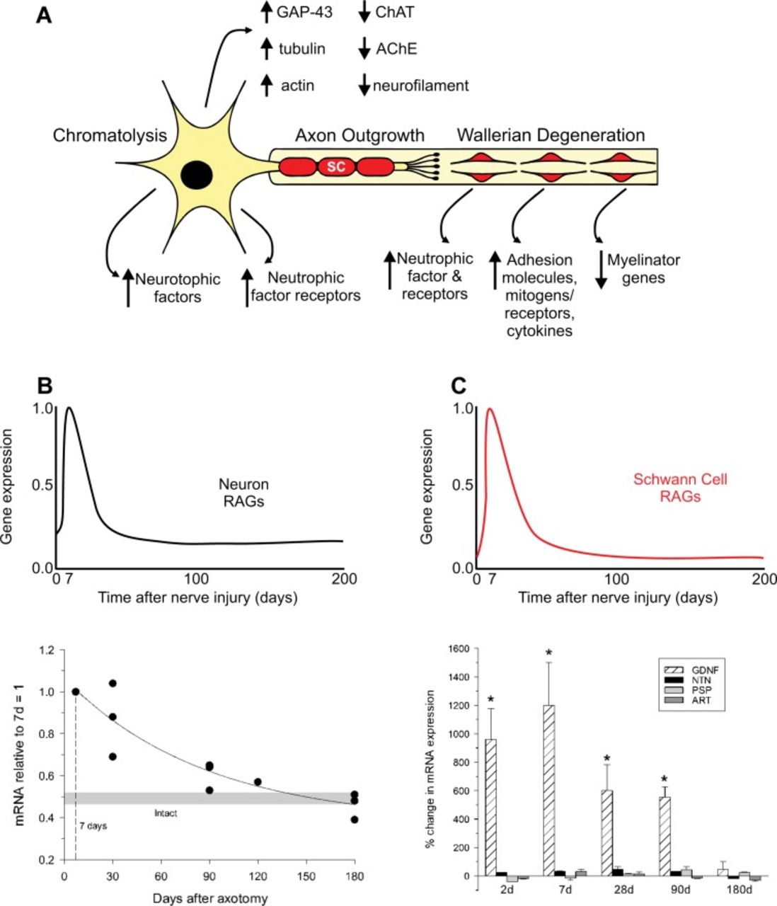
  • Figure 2.
    • Download figure
    • Open in new tab
    • Download powerpoint
    Figure 2.

    After nerve injury, regeneration-associated genes (RAGs) are upregulated transiently in the neurons while genes associated with normal synaptic transmission are downregulated (A and B). Schwann cells in the denervated nerve stumps undergo proliferation during Wallerian degeneration and express many RAGs while myelin-associated genes are downregulated (A). The gene profiles support the outgrowth of axons, but the expression is very short lived such that over time (while regenerating axons grow at a slow rate of 1 mm/d) the expression of RAGs is downregulated and the capacity of injured neurons to regenerate their axons and Schwann cells to support regeneration is diminished (C). Examples of progressive decline in mRNA levels are plotted for tubulin in neurons and GDNF in Schwann cells in the graphs.
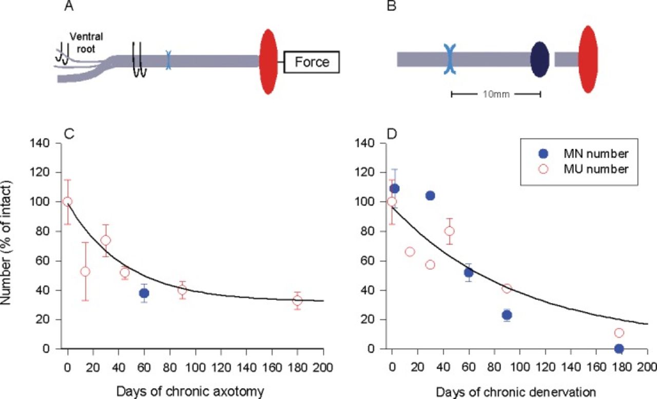
  • Figure 3.
    • Download figure
    • Open in new tab
    • Download powerpoint
    Figure 3.

    Regenerative capacity declines with time due to prolonged axotomy and Schwann cell denervation. In rats, we experimentally prolonged either (A) the duration of time during which motoneurons were prevented from regenerating their axons (chronic axotomy; by delaying the suture of the proximal nerve stump to a freshly denervated distal nerve stump) or (B) the denervation of Schwann cells in the distal nerve stumps (chronic denervation; by delaying the suture of a freshly cut proximal nerve stump to a nerve stump that was chronically denervated prior to nerve repair). The capacity of the neurons for regeneration after chronic axotomy of motoneurons or chronic denervation of Schwann cells and target muscles was determined either by (a) calculating the number of reinnervated muscle units (the nerve and the muscle fibers that the one motoneuron supplies) using force measurements in response to stimulation of single axons and the muscle nerve or (b) counting backlabelling motoneurons that had regenerated their axons successfully by application of a retrograde dye to the regenerating axons in the nerve stump distal to the site of nerve repair. The evaluations of regenerative success obtained by the methods of motoneuron counts and counts of motor units were in good agreement demonstrating the progressive decline in regenerative capacity as a function of (C) chronic axotomy of the motoneurons and (D) chronic denervation of the Schwann cells.

PreviousNext
Back to top

In this issue

Ochsner Journal
Vol. 13, Issue 1
Mar 2013
  • Table of Contents
  • Index by author
Print
Download PDF
Email Article

Thank you for your interest in spreading the word on Ochsner Journal.

NOTE: We only request your email address so that the person you are recommending the page to knows that you wanted them to see it, and that it is not junk mail. We do not capture any email address.

Enter multiple addresses on separate lines or separate them with commas.
Neurobiology of Peripheral Nerve Injury, Regeneration, and Functional Recovery: From Bench Top Research to Bedside Application
(Your Name) has sent you a message from Ochsner Journal
(Your Name) thought you would like to see the Ochsner Journal web site.
CAPTCHA
This question is for testing whether or not you are a human visitor and to prevent automated spam submissions.
Citation Tools
Neurobiology of Peripheral Nerve Injury, Regeneration, and Functional Recovery: From Bench Top Research to Bedside Application
Wale Sulaiman, Tessa Gordon
Ochsner Journal Mar 2013, 13 (1) 100-108;

Citation Manager Formats

  • BibTeX
  • Bookends
  • EasyBib
  • EndNote (tagged)
  • EndNote 8 (xml)
  • Medlars
  • Mendeley
  • Papers
  • RefWorks Tagged
  • Ref Manager
  • RIS
  • Zotero
Share
Neurobiology of Peripheral Nerve Injury, Regeneration, and Functional Recovery: From Bench Top Research to Bedside Application
Wale Sulaiman, Tessa Gordon
Ochsner Journal Mar 2013, 13 (1) 100-108;
del.icio.us logo Twitter logo Facebook logo Mendeley logo
  • Tweet Widget
  • Facebook Like
  • Google Plus One

Jump to section

  • Article
    • ABSTRACT
    • INTRODUCTION
    • REGENERATIVE RESPONSE AFTER NERVE INJURY AND ITS ROLE IN FUNCTIONAL OUTCOME
    • EXPERIMENTAL PARADIGMS AND ASSESSMENT OF AXONAL REGENERATION AFTER NERVE INJURY AND MICROSURGICAL REPAIR
    • CONCLUSIONS
    • Footnotes
    • REFERENCES
  • Figures & Data
  • References
  • Info & Metrics
  • PDF

Cited By...

  • Recovery of retinal terminal fields after traumatic brain injury: evidence of collateral sprouting and sexual dimorphism
  • Evaluation of distal facial nerve branches contribution to facial nerve paralysis in rodents
  • Schwann Cells as Orchestrators of Nerve Repair: Implications for Tissue Regeneration and Pathologies
  • Nerve injury converts Schwann cells in a persisting repair state in human neuroma tissue
  • Targeting Vasohibins to Promote Axon Regeneration
  • Human Motor Endplate Survival after Chronic Peripheral Nerve Injury
  • VASH1/2 inhibition accelerates functional recovery of injured nerves
  • Angiogenesis is critical for the regenerative effects of exercise
  • Repetitive Peripheral Magnetic Stimulation (rPMS) in Subjects With Lumbar Radiculopathy: An Electromyography-guided Prospective, Randomized Study
  • Transforming Growth Factor Beta 1 Regulates Fibroblast Growth Factor 7 Expression in Schwann Cells
  • Collagen XIII Is Required for Neuromuscular Synapse Regeneration and Functional Recovery after Peripheral Nerve Injury
  • After Nerve Injury, Lineage Tracing Shows That Myelin and Remak Schwann Cells Elongate Extensively and Branch to Form Repair Schwann Cells, Which Shorten Radically on Remyelination
  • Nerve injuries of the upper extremity and hand
  • STAT3 Controls the Long-Term Survival and Phenotype of Repair Schwann Cells during Nerve Regeneration
  • Recovery of erectile function comparing autologous nerve grafts, unseeded conduits, Schwann-cell-seeded guidance tubes and GDNF-overexpressing Schwann cell grafts
  • Recent Publications by Ochsner Authors
  • Google Scholar

More in this TOC Section

  • Rhabdomyolysis: Pathogenesis, Diagnosis, and Treatment
  • iPhone and iPad Use in Orthopedic Surgery
  • Drug-Induced Acute Pancreatitis: A Review
Show more Reviews and Commentaries

Similar Articles

Keywords

  • Axotomy
  • cytokines
  • denervation
  • nerve growth factors
  • nerve regeneration
  • neurobiology
  • peripheral nerve injuries
  • recovery of function
  • Schwann cells

Ochsner Journal Blog

Current Post

Ochsner Journal Features Reports from the BioDesign Lab and Pediatric Cardiology

Our Content

  • Home
  • Current Issue
  • Ahead of Print
  • Archive
  • Featured Contributors
  • Ochsner Journal Blog
  • Archive at PubMed Central

Information & Forms

  • Instructions for Authors
  • Instructions for Reviewers
  • Submission Checklist
  • FAQ
  • License for Publishing-Author Attestation
  • Patient Consent Form
  • Submit a Manuscript

Services & Contacts

  • Permissions
  • Sign up for our electronic table of contents
  • Feedback Form
  • Contact Us

About Us

  • Editorial Board
  • About the Ochsner Journal
  • Ochsner Health
  • University of Queensland-Ochsner Clinical School
  • Alliance of Independent Academic Medical Centers

© 2025 Ochsner Clinic Foundation

Powered by HighWire