
Main menu

  • Home
  • Content
    • Current
    • Ahead of print
    • Archive
  • Info for
    • Authors
    • Reviewers
  • About Us
    • About the Ochsner Journal
    • Editorial Board
  • More
    • Alerts
    • Feedback
  • Other Publications
    • Ochsner Journal Blog

User menu

  • My alerts
  • Log in

Search

  • Advanced search
Ochsner Journal
  • Other Publications
    • Ochsner Journal Blog
  • My alerts
  • Log in
Ochsner Journal

Advanced Search

  • Home
  • Content
    • Current
    • Ahead of print
    • Archive
  • Info for
    • Authors
    • Reviewers
  • About Us
    • About the Ochsner Journal
    • Editorial Board
  • More
    • Alerts
    • Feedback
Review ArticleReviews and Commentaries

Diabetic Nephropathy: Lessons From the Mouse

Himanshu Vashistha and Leonard Meggs
Ochsner Journal March 2013, 13 (1) 140-146;
Himanshu Vashistha
*Nephrology Research Laboratory, Institute of Translational Research, and
PhD
  • Find this author on Google Scholar
  • Find this author on PubMed
  • Search for this author on this site
Leonard Meggs
†Department of Nephrology, Ochsner Clinic Foundation, New Orleans, LA
MD
  • Find this author on Google Scholar
  • Find this author on PubMed
  • Search for this author on this site
  • Article
  • Figures & Data
  • References
  • Info & Metrics
  • PDF
Loading

Article Figures & Data

Figures

  • Figure 1.
    • Download figure
    • Open in new tab
    • Download powerpoint
    Figure 1.

    Light micrographs showing periodic acid-Schiff staining of glomeruli counterstained with hematoxylin from Akita (Ins2+/C96Y/B2R+/+) and Akita (Ins2+/C96Y/B2R-/-) genotypes. B2R, bradykinin 2 receptor.

  • Figure 2.
    • Download figure
    • Open in new tab
    • Download powerpoint
    Figure 2.

    Modular organization of ShcA isoforms. CH, collagen-homologous; PTB, phosphotyrosine binding domain; SH, Src homology.
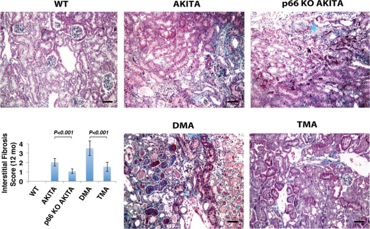
  • Figure 3.
    • Download figure
    • Open in new tab
    • Download powerpoint
    Figure 3.

    Periodic acid-Schiff (PAS) staining of kidney sections. The Ins2 mutation alone increases accumulation of PAS-positive matrix at 12 months of age, which is enhanced by the absence of the bradykinin 2 receptor. The p66 null mutation attenuates renal histological changes at corresponding intervals: nodular PAS-positive matrix (black arrows in Akita and double mutant Akita [DMA]), mesangiolysis (yellow arrows in Akita and DMA), tubular dilation (blue arrow in DMA), and microaneurysm (arrowhead in DMA). The scale bar is 10 μm. The insert shows the histologic analysis of glomerulosclerosis by semiquantitative morphometric analysis. Results are presented as mean ± standard deviation; n=5 in each group; P<0.01 Akita (Ins2+/C96Y) vs p66 knockout (KO) Akita (p66-/-/Ins2+/C96Y); P<0.001 DMA (Ins2+/C96Y/B2R-/-) vs triple mutant Akita (TMA) (p66-/-/Ins2+/C96Y/B2R-/-). WT, wild type.
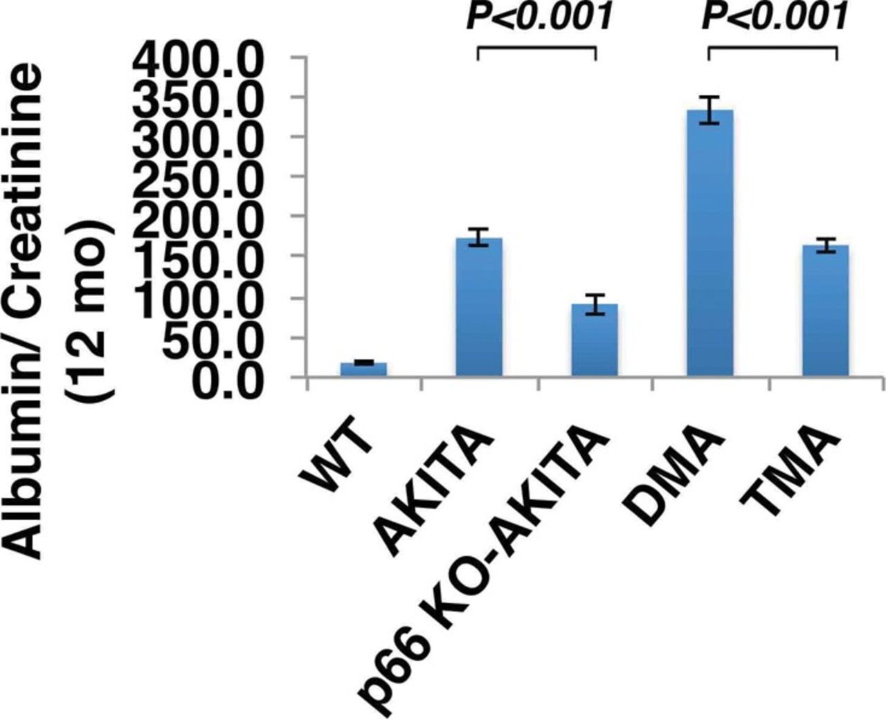
  • Figure 4.
    • Download figure
    • Open in new tab
    • Download powerpoint
    Figure 4.

    The p66 null mutation attenuates urine albumin excretion. Results are presented as mean ± standard deviation; n=5 in each group. P<0.001 Akita (Ins2+/C96Y) vs p66 knockout (KO) Akita (p66-/-/Ins2+/C96Y); P<0.001 double mutant Akita (DMA) (Ins2+/C96Y/B2R-/-) vs triple mutant Akita (TMA) (p66-/-/Ins2+/C96Y/B2R-/-).

  • Figure 5.
    • Download figure
    • Open in new tab
    • Download powerpoint
    Figure 5.

    Trichrome staining of the renal cortex. Double mutant Akita (DMA) mice (Ins2+/C96Y/B2R-/-) show increased interstitial fibrosis at 12 months of age, whereas the p66 null mutation attenuates interstitial fibrosis at corresponding intervals. The scale bar is 50 μm. The insert shows the histologic analysis of interstitial fibrosis assessed by semiquantitative evaluation. Results are presented as mean ± standard deviation; n=5 in each group. P<0.001 Akita vs p66 knockout (KO) Akita; P<0.001 DMA vs triple mutant Akita (TMA) (p66-/-/Ins2+/C96Y/B2R-/-). WT, wild type.
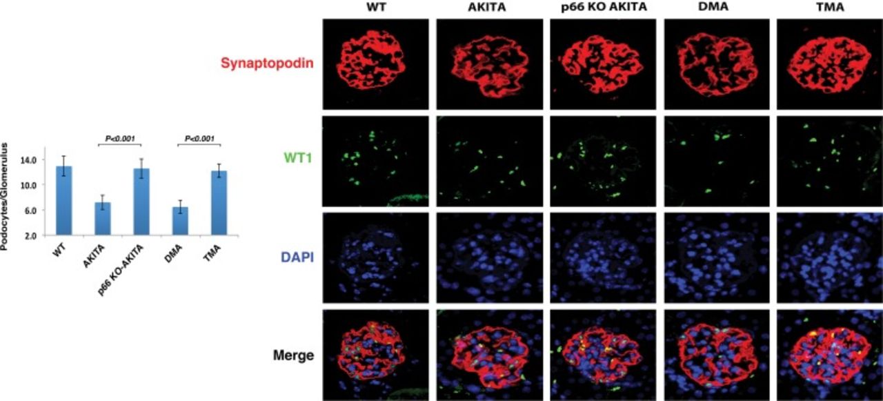
  • Figure 6.
    • Download figure
    • Open in new tab
    • Download powerpoint
    Figure 6.

    Podocyte number/glomerulus. WT1 (Wilms tumor 1) is expressed in the podocyte nucleus only. Marker-expressing nuclei were counted in the kidneys of Akita mice. Results are presented as mean ± standard deviation; n=5 in each group. Thirty glomeruli from each mouse were analyzed; P<0.001 p66 knockout (KO) Akita (p66-/-/Ins2+/C96Y) vs Akita (Ins2+/C96Y); P<0.001 double mutant Akita (DMA) (Ins2+/C96Y/B2R-/-) vs triple mutant Akita (TMA) (p66-/-/Ins2+/C96Y/B2R-/-). Confocal images of glomeruli show the podocyte-specific marker synaptopodin (red), podocyte nuclear marker WT1 (green), and nuclear counterstain DAPI (blue) in kidney sections at 12 months of age. Merge shows the expression of synaptopodin, WT1, and DAPI.

  • Figure 7.
    • Download figure
    • Open in new tab
    • Download powerpoint
    Figure 7.

    (A) Representative immunoblot analysis of ShcA isoforms in conditionally immortalized differentiated human podocytes (CIDHPs) stably transfected with p66 shRNA. (B) CIDHP/p66 shRNA shows inhibition of HG-induced reactive oxygen species (ROS) metabolism. (C) Quantitative analysis of ROS. (D) CIDHP/p66 shRNA shows inhibition of HG-induced apoptosis signal, *P<0.01. EV-CIDHP, empty vector-transduced conditionally immortalized differentiated human podocytes; HG, high glucose (40 mM); Manni, mannitol; NG, normal glucose (5 mM); OD, optical density.

  • Figure 8.
    • Download figure
    • Open in new tab
    • Download powerpoint
    Figure 8.

    Representative immunoblot analysis of sirtuin 1 (SIRT1) deacetylase and acetylation of lysine-382 of p53 protein. SIRT1 expression is downregulated in Akita diabetic mice but restored in p66 knockout mice. Similarly, the expression of acetyl p53 is upregulated in Akita diabetic mice but returns to normal expression levels in p66 knockout mice. β-actin was used as a loading control. KO, knockout; WT, wild type.

PreviousNext
Back to top

In this issue

Ochsner Journal
Vol. 13, Issue 1
Mar 2013
  • Table of Contents
  • Index by author
Print
Download PDF
Email Article

Thank you for your interest in spreading the word on Ochsner Journal.

NOTE: We only request your email address so that the person you are recommending the page to knows that you wanted them to see it, and that it is not junk mail. We do not capture any email address.

Enter multiple addresses on separate lines or separate them with commas.
Diabetic Nephropathy: Lessons From the Mouse
(Your Name) has sent you a message from Ochsner Journal
(Your Name) thought you would like to see the Ochsner Journal web site.
CAPTCHA
This question is for testing whether or not you are a human visitor and to prevent automated spam submissions.
Citation Tools
Diabetic Nephropathy: Lessons From the Mouse
Himanshu Vashistha, Leonard Meggs
Ochsner Journal Mar 2013, 13 (1) 140-146;

Citation Manager Formats

  • BibTeX
  • Bookends
  • EasyBib
  • EndNote (tagged)
  • EndNote 8 (xml)
  • Medlars
  • Mendeley
  • Papers
  • RefWorks Tagged
  • Ref Manager
  • RIS
  • Zotero
Share
Diabetic Nephropathy: Lessons From the Mouse
Himanshu Vashistha, Leonard Meggs
Ochsner Journal Mar 2013, 13 (1) 140-146;
del.icio.us logo Twitter logo Facebook logo Mendeley logo
  • Tweet Widget
  • Facebook Like
  • Google Plus One

Jump to section

  • Article
    • ABSTRACT
    • INTRODUCTION
    • MICE MODELS
    • OUR RESEARCH
    • CONCLUSION
    • Footnotes
    • REFERENCES
  • Figures & Data
  • References
  • Info & Metrics
  • PDF

Cited By...

  • Recent Publications by Ochsner Authors
  • Google Scholar

More in this TOC Section

  • Drug-Induced Acute Pancreatitis: A Review
  • Rhabdomyolysis: Pathogenesis, Diagnosis, and Treatment
  • iPhone and iPad Use in Orthopedic Surgery
Show more Reviews and Commentaries

Similar Articles

Keywords

  • diabetic nephropathies
  • oxidative stress
  • p66Shc protein
  • proteinuria

Our Content

  • Home
  • Current Issue
  • Ahead of Print
  • Archive
  • Featured Contributors
  • Ochsner Journal Blog
  • Archive at PubMed Central

Information & Forms

  • Instructions for Authors
  • Instructions for Reviewers
  • Submission Checklist
  • FAQ
  • License for Publishing-Author Attestation
  • Patient Consent Form
  • Submit a Manuscript

Services & Contacts

  • Permissions
  • Sign up for our electronic table of contents
  • Feedback Form
  • Contact Us

About Us

  • Editorial Board
  • About the Ochsner Journal
  • Ochsner Health
  • University of Queensland-Ochsner Clinical School
  • Alliance of Independent Academic Medical Centers

© 2026 Ochsner Clinic Foundation

Powered by HighWire