
Abstract
Background Myelomeningocele is the most common form of congenital central nervous system defect that is compatible with life. Most patients with myelomeningocele have significant functional impairment of ambulation and bowel and bladder function, require permanent cerebrospinal fluid diversion with shunting, and have significant morbidity and mortality from hindbrain herniation (Chiari II malformation). The advent of intrauterine surgery has provided new opportunities to better address this lifelong debilitating disease.
Case Report The patient was a 19-year-old gravida 2 para 1 at 22-6/7 weeks whose fetus was diagnosed with an open neural tube defect and further demonstrated to have ventriculomegaly and hindbrain herniation. Amniocentesis confirmed normal karyotype and the presence of acetylcholinesterase. After an intrauterine procedure, the patient underwent cesarean section at 35-5/7 weeks and delivered a male infant. His spinal incision was well healed at birth without any evidence of cerebrospinal fluid leakage, and his extremities were normal in appearance, range of motion, and movement. The infant also has maintained relatively normal, age-appropriate bowel and bladder function and has no obvious neurologic deficit.
Conclusion As the benefit of fetal surgery becomes more widely accepted, quality of care and patient safety must be at the forefront of any institution's effort to offer fetal surgery. Given the current prevalence of spina bifida and the amount of resources required to treat this disease effectively either in utero or postnatally, it is our opinion that the treatment of spina bifida should be regionalized to tertiary referral centers with the interdisciplinary expertise to offer comprehensive treatment for all aspects of the disease and all phases of care for the patients.
- Arnold-Chiari malformation
- fetal therapies
- hydrocephalus
- hysterotomy
- meningomyelocele
- spinal dysraphism
- ventriculoperitoneal shunt
INTRODUCTION
Myelomeningocele, or herniation of the neural elements through a posterior spinal and skin defect, results from failure of the neural tube to close during the early period of embryonic development. Myelomeningocele's incidence has decreased to less than 1 per 1,000 live births since the advent of folic acid supplementation,1 but it still remains a formidable disease from a patient care as well as healthcare cost perspective. It is the most common form of congenital central nervous system defect compatible with life, and its associated symptoms are a lifelong medical burden for affected patients and their families. The defect has traditionally been treated postpartum with operative repair within 72 hours of birth and with early multidisciplinary therapy. Despite aggressive surgical and medical treatment, more than 80% of patients require ventriculoperitoneal shunting (VPS) and have ambulation impairments such that two-thirds of patients are wheelchair bound by adulthood. Virtually all myelomeningocele patients have significant hindbrain herniation, also known as Chiari II malformation, that is believed to contribute to premature death in this patient population.
ESTABLISHMENT OF INTRAUTERINE MYELOMENINGOCELE REPAIR
The concept of in utero surgery is not a novel idea; it was first performed in dogs in the 1930s2 and advanced further by reducing amniotic fluid loss in the 1960s.3 In 1980 at the University of California, San Francisco (UCSF), Harrison et al developed a successful modern animal model for fetal surgery.4 Results of animal models and surgeries for spina bifida in the 1980s established technical feasibility and showed early evidence of improved outcomes.5,6 Fetal surgery has been employed for various congenital diseases including congenital cystic adenomatoid malformation of the lung,7 congenital diaphragmatic hernia,8 sacrococcygeal teratoma,9 and fetal airway obstruction.10
The hypothesis that intrauterine repair of myelomeningocele would improve outcomes is based on evidence that exposure of neural elements to the amniotic fluid and the intrauterine environment results in secondary injuries to the neural elements and facilitates hindbrain herniation. Therefore, earlier closure may prevent secondary injuries and result in improved functional outcomes. The first fetal surgeries for spina bifida repair in animals were performed in 199411,12 and were followed by successful repairs in humans in 1997 and 1998 at Vanderbilt University Medical Center and Children's Hospital of Philadelphia (CHOP), respectively.13-15
Intrauterine surgery is not without significant ethical implications and controversies. Lawyers, ethicists, doctors, nurses, and the media at large have discussed the implications of intrauterine surgery since the 1980s,16-19 particularly as it relates to the mother's autonomy in making healthcare decisions, as maternal complication is a risk to be weighed against the potential benefit of improving the fetus's outcome. Two of the most frequent complications of hysterotomy are wound dehiscence and uterine scarring that may complicate future pregnancy. Another major potential complication is preterm labor that can result in an unviable fetus, particularly if delivery occurs before the 24th week. Regardless, most mothers and physicians feel the benefits of improved outcomes outweigh the ethical and moral considerations of intrauterine fetal surgery, so fetal repairs have been performed with gradually increasing frequency since the 1990s. However, a lack of sufficient data prevented widespread establishment of the surgery.
After promising initial data20-22 and with international medical consensus, the National Institutes of Health–funded randomized prospective Management of Myelomeningocele Study (MOMS) trial was initiated in 2003 at 3 tertiary centers: UCSF, CHOP, and Vanderbilt. While the study was active, all other centers throughout the country suspended in utero myelomeningocele repairs. To be eligible to participate, women had to have a singleton pregnancy with a fetus meeting the following requirements: a lesion with the upper end located between T1 and S1, evidence of hindbrain herniation, gestational age of 19 to 25.9 weeks, and a normal karyotype. Participants were also required to be at least 18 years old, be US residents, have a body mass index <35, have no previous hysterotomy, and have no other apparent risk factor for preterm birth.23
The 2 primary outcomes of the study were (1) a composite of fetal/neonatal death or the need for a cerebrospinal fluid (CSF)-diverting shunt at 12 months and (2) a composite score of development and motor function at 30 months. Secondary outcomes were defined for the fetus/infant and the mother. For infants, secondary outcomes included radiographic appearance of Chiari II malformation, time to eligibility for shunt placement, locomotion, motor and developmental outcome measures, functional examination, and degree of disability. Secondary outcomes for mothers focused on surgical and pregnancy complications.
Because of favorable primary and secondary outcomes, the trial was terminated early to allow more patients to have prenatal surgery. For the first primary outcome, only 68% in the prenatal group compared with 98% in the postnatal group (P<0.001) had fetal or neonatal death or met criteria for a VPS. Only 40% of prenatal patients met criteria for a shunt, whereas 82% of postnatal patients did. For the second primary outcome, composite functional measures were also better in the prenatal surgery group (P=0.007) than in the postnatal group.
Pregnancy-related outcomes were worse for mothers in the prenatal group; they were more likely to suffer poor-quality wound healing or wound dehiscence, chorioamniotic separation, placental abruption, and spontaneous membrane rupture. Fetuses from the prenatal group were more likely to be delivered prematurely, at an average age of 34.1 weeks as opposed to 37.3 weeks in the postnatal group. Prenatal-surgery infants were also more likely to require further procedures for delayed spinal cord tethering.
Secondary outcomes were also more favorable in the prenatal-surgery infants compared to the postnatal group. Of the prenatal group, 36% had no evidence of hindbrain herniation as opposed to 4% in the postnatal group. Level of function was more likely to be 2 or more levels better than expected according to the level of the lesion (32% vs 12%, P=0.005) and less likely to be 2 or more levels worse (13% vs 28%, P=0.03) in the prenatal vs postnatal patients. Prenatal patients were also more likely to be able to walk without prosthetics and orthotics than their postnatal counterparts (42% vs 21%, P=0.01). Cognition was equal in the 2 groups; however, motor function on both the Bayley and Peabody motor scales and disability as measured by the Functional Independence Measure instrument showed better outcomes in the prenatal surgery group.
The overall results are compelling, given a relative risk reduction (RRR) of 51% with respect to shunting; an RRR of 33% with respect to hindbrain herniation; and better outcomes with respect to ambulation, motor function, and disability function. These advantages are not without risks, as significantly more prenatal patients were born preterm, and mothers undergoing hysterotomy are more likely to have complications from scarring that might affect future pregnancies. Unfortunately, in utero repair has not conferred an advantage with respect to infants' bladder function.24
THE OCHSNER EXPERIENCE
Ochsner has a long and successful track record of treating and caring for children with spina bifida. After the publication of the MOMS data, we felt that it was imperative for Ochsner to be able to offer families this option. We were able to assemble a fetal surgery team with active participation of pediatric neurosurgery, maternal fetal medicine, pediatric surgery, obstetric anesthesia, pediatric cardiology, and pediatrics. After more than a year of preparation, the first fetal surgery in the southeast region was performed on December 3, 2012.
We have thus far performed 6 fetal repairs without any intraoperative complications, although a major shortcoming of our current experience is a limited follow-up period. We are also concurrently following 4 infants locally who had fetal repairs at Vanderbilt and CHOP. We present our first patient as an illustrative case because we have the longest follow-up for this patient and her child.
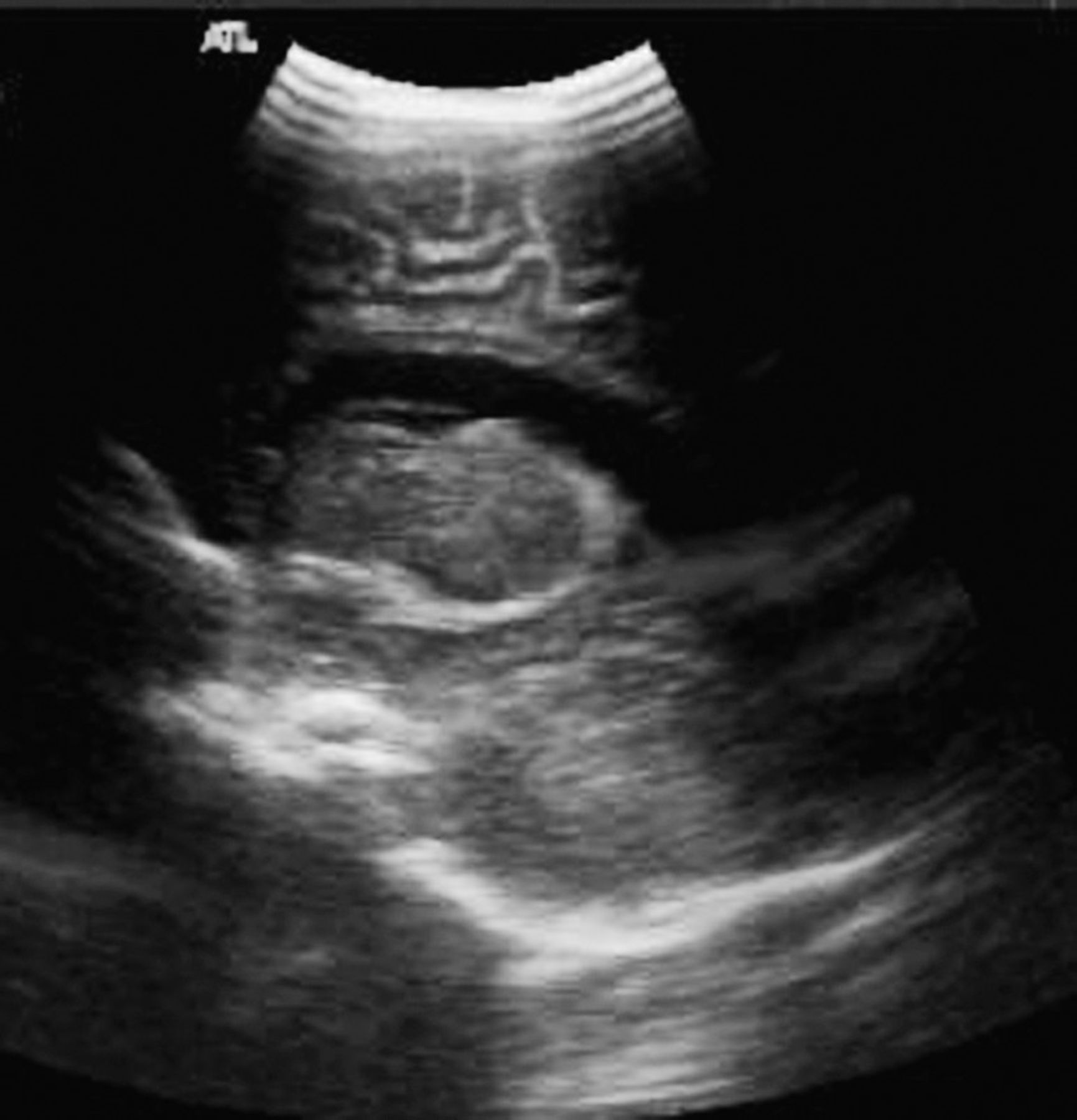
The patient was a 19-year-old gravida 2 para 1 at 22-6/7 weeks whose fetus was diagnosed with an open neural tube defect by ultrasound that was confirmed with maternal magnetic resonance imaging (MRI). Ultrasound and MRI further demonstrated ventriculomegaly (Figure 1) and hindbrain herniation (Chiari II malformation). Amniocentesis confirmed normal karyotype and the presence of acetylcholinesterase. After full evaluation and counseling by the Ochsner fetal surgery team, the patient and her family wished to proceed with intrauterine repair.

In utero ultrasound (A) and magnetic resonance imaging (B) demonstrate open neural tube defect and (C) marked ventriculomegaly.
The surgical technique employed was the standard as established by the MOMS investigators, with a minor modification: we included Seprafilm (Sanofi US) under the dural repair to help minimize the incidence of spinal cord tethering (Figure 2). The patient was maintained postoperatively on magnesium sulfate drip for several days with an unremarkable hospital course and was discharged home on postoperative day 7 with plans to return for elective cesarean section (C-section) at 36 weeks.

Photographs from the operating room demonstrating (A) exposure of the myelomeningocele through the hysterotomy, (B) placement of Seprafilm during closure of the myelomeningocele, (C) the repaired myelomeningocele defect, and (D) the hysterotomy repair.
At 35-5/7 weeks, she presented with uterine contractions and underwent C-section. She delivered a male infant with Apgar scores of 8 and 8. His spinal incision was well healed at birth without any evidence of CSF leakage. Both lower extremities were normal in appearance, range of motion, and movement. Head ultrasound at birth (Figure 3) noted only mild prominence of the ventricular system and showed improvement when compared with preoperative in utero imaging.

Echoencephalography demonstrating the lateral ventricle on day 1 of life.
The infant has maintained relatively normal bladder function with mild constipation. At 7 months of age, he has continued to do well without any obvious neurologic deficit and demonstrates normal leg movement, age-appropriate bladder function, and excellent cosmetic results. On follow-up echoencephalography when the infant was 4 months old, we found stability of the lateral ventricle without any evidence of hydrocephalus (Figure 4) and no obvious hindbrain herniation.
Echoencephalography demonstrating stability of the lateral ventricle at 4 months of life.
DISCUSSION
Intrauterine repair of myelomeningocele confers multiple advantages to infants, including lower rates of shunt dependency, lower rates of hindbrain herniation, and better motor and disability functional outcomes. Our first infant repaired in utero appears to have favorable outcomes in all the aforementioned areas. Given the high rate of shunt failure, the healthcare issues related to being wheelchair bound, and the complications from hindbrain herniation, the long-term healthcare benefits and savings may be immeasurable. Although we have performed 6 successful repairs, our current follow-up is still limited and it is too early to know our true long-term efficacy rate and whether we can reproduce the favorable outcomes seen with the MOMS trial.
Fetal surgery is a challenging, and at times controversial, medical technology that has shown tremendous progress in the past few decades. In addition to spina bifida, fetal surgery may benefit myriad other congenital abnormalities. Beyond the immediate possible medical benefits to individual patients, the tremendous downstream healthcare savings opportunities for patients, their families, and the healthcare system at large cannot be overlooked. A study of the fiscal cost of myelomeningocele has suggested that each patient with spina bifida spends a mean of $10,000 to $41,000 (2003 dollars) more per year in healthcare than the average person without spina bifida.25 As spina bifida patients are living longer and have a more normal life expectancy, the lifetime cost-saving potentials offered by in utero repair are great. Yet these benefits must be weighed against potential surgical risks, the most significant of which is preterm delivery. Follow-up data are too limited to indicate if long-term adverse risks to the child or mother exist in preterm surgery cases.
CONCLUSION
Assembly of a fetal surgery team is both expertise- and resource-intensive and requires superlative focus of all involved subspecialties on both quality and safety. As the benefit of fetal surgery becomes more widely accepted, quality of care and patient safety must be at the forefront of any institution's effort to offer fetal surgery. Given the current prevalence of spina bifida and the amount of resources required to treat this disease effectively either in utero or postnatally, it is our opinion that the treatment of spina bifida should be regionalized to tertiary referral centers with the interdisciplinary expertise to offer comprehensive treatment for all aspects of the disease and all phases of care for the patients. These centers will have sufficient volume to deliver the highest quality care in the most cost-effective manner.
This article meets the Accreditation Council for Graduate Medical Education and the American Board of Medical Specialties Maintenance of Certification competencies for Patient Care, Medical Knowledge, and Systems-Based Practice.
Footnotes
The authors have no financial or proprietary interest in the subject matter of this article.
- © Academic Division of Ochsner Clinic Foundation


