
Main menu

  • Home
  • Content
    • Current
    • Ahead of print
    • Archive
  • Info for
    • Authors
    • Reviewers
  • About Us
    • About the Ochsner Journal
    • Editorial Board
  • More
    • Alerts
    • Feedback
  • Other Publications
    • Ochsner Journal Blog

User menu

  • My alerts
  • Log in

Search

  • Advanced search
Ochsner Journal
  • Other Publications
    • Ochsner Journal Blog
  • My alerts
  • Log in
Ochsner Journal

Advanced Search

  • Home
  • Content
    • Current
    • Ahead of print
    • Archive
  • Info for
    • Authors
    • Reviewers
  • About Us
    • About the Ochsner Journal
    • Editorial Board
  • More
    • Alerts
    • Feedback
Case ReportCase Reports

Primary Amyloidosis of the Breast Presenting as a Solitary Nodule: Case Report and Review of the Literature

Mary E. Huerter, Rasheed Hammadeh, Quan Zhou, Asher Weisberg and Adam I. Riker
Ochsner Journal June 2014, 14 (2) 282-286;
Mary E. Huerter
1Department of Surgery, University of Illinois at Chicago, Chicago, IL
MD
  • Find this author on Google Scholar
  • Find this author on PubMed
  • Search for this author on this site
Rasheed Hammadeh
2Department of Pathology, Advocate Cancer Institute, Advocate Christ Medical Center, Oak Lawn, IL
MD
  • Find this author on Google Scholar
  • Find this author on PubMed
  • Search for this author on this site
Quan Zhou
3Department of Pathology, Beijing Shijitan Hospital, Capital Medical University, Beijing, China
MD
  • Find this author on Google Scholar
  • Find this author on PubMed
  • Search for this author on this site
Asher Weisberg
1Department of Surgery, University of Illinois at Chicago, Chicago, IL
BS
  • Find this author on Google Scholar
  • Find this author on PubMed
  • Search for this author on this site
Adam I. Riker
4Department of Surgery, Advocate Cancer Institute, Advocate Christ Medical Center, Oak Lawn, IL
MD, FACS
  • Find this author on Google Scholar
  • Find this author on PubMed
  • Search for this author on this site
  • Article
  • Figures & Data
  • References
  • Info & Metrics
  • PDF
Loading

Abstract

Background Amyloidosis of the breast is an unusual diagnosis. Overall, only 2 major case series have been published in the literature since the time amyloidosis was first reported in 1973, highlighting the rare nature of this disease. Clinically, the patient may present with a breast mass or simply increased breast density and skin thickening. As such, amyloidosis may mimic the appearance of a number of pathologies, both benign and malignant.

Case Report We report a single case of a patient who presented with a breast mass and was ultimately diagnosed with primary amyloidosis of the breast. We also present a review of the literature to date.

Conclusion The main treatment for primary amyloidosis of the breast is surgical removal, most often accomplished with breast conservation via lumpectomy. In performing the lumpectomy, the goal should be to obtain negative surgical margins; however, no strong data exist to suggest that such lesions would recur with a microscopically positive margin.

Keywords
  • Amyloidosis
  • Congo red
  • mammogram
  • mastectomy–segmental

INTRODUCTION

Amyloidosis of the breast is a rare entity, with only 2 major case series reported in the nearly 40 years since it was first reported in 1973.1 Amyloidosis is the extracellular deposition of insoluble beta-pleated fibrillar proteins.2 It may be localized or systemic, with the isolated involvement of the breast seldom reported. Staining for amyloid protein with Congo red reveals a characteristic apple-green birefringence under polarized light.3 Amyloidosis of the breast can distribute in a periductal, perivascular, or intralobar pattern; can create a foreign body–like reaction with infiltration of multinucleated giant cells; and can have an affinity for calcium.2 Clinically, the patient may present with a breast mass or simply with increased breast density and skin thickening.4 Radiographic imaging reveals a wide variety of mammographic and ultrasound findings, including similar patterns of ductal carcinoma in situ and fibrocystic change.4 As such, amyloidosis may mimic the appearance of a number of pathologies, both benign and malignant. However, more commonly, amyloidosis of the breast is a manifestation of an overall systemic disorder coexisting in the presence of systemic amyloidosis or a hematologic malignancy, mandating a thorough evaluation of the patient for these disorders.5,6

CASE REPORT

A 52-year-old African American female undergoing her regularly scheduled screening mammogram was found to have a new questionable area of focal asymmetry within the left breast. The mammogram was given an overall Breast Imaging-Reporting and Data System (BI-RADS) category 3 assessment (on a scale of 0-6), with a 6-month follow-up of this area recommended. The follow-up mammogram revealed an evolving mass in the same area, measuring 2.0 × 3.0 × 1.5 cm by both mammogram and ultrasound and considered a BI-RADS category 4 abnormality with biopsy recommended (Figure 1). The patient underwent an ultrasound-guided core biopsy, with the final pathology revealing amyloidosis with a lymphoplasmacytic infiltrate surrounding the breast ducts and some blood vessels.

Figure 1.
  • Download figure
  • Open in new tab
  • Download powerpoint
Figure 1.

A: Mammogram with craniocaudal view of the breast with needle guidewire in place just prior to operative procedure. The previously placed core biopsy clip is visible within the mass. B: Medial-lateral view of the breast with the needle guidewire placed. C: Imaged lumpectomy specimen performed intraoperatively, showing complete gross removal of the mass, core biopsy clip, and guidewire.

Initial staining for amyloid protein was performed with Congo red dye and revealed a characteristic apple-green birefringence that represents a positive finding for amyloid within the specimen (Figure 2). No evidence was found of breast cancer or other breast pathology; the associated immunohistochemical analysis revealed both kappa and lambda staining (10:1 kappa to lambda ratio). The patient was evaluated first by a medical oncologist, and a series of studies did not reveal any significant findings of amyloidosis. Bone marrow biopsy revealed normocellular bone marrow that was negative for plasma cell dyscrasia on flow cytometric analysis and negative for the presence of amyloid. Serum protein electrophoresis revealed a polyclonal increase in the gamma region. With no clinical symptoms reported and no evidence of systemic amyloidosis, the patient was seen by a surgical oncologist for further evaluation and possible removal of this breast mass. The patient subsequently underwent a needle-localized lumpectomy of this mass (Figure 3), and the final pathology revealed an amyloidoma with surrounding margins of breast parenchyma containing interstitial fibrosis, fibrocystic change, and periductal chronic inflammation (Figure 4). No evidence of malignancy was found and all of the surgical margins were negative.

Figure 2.
  • Download figure
  • Open in new tab
  • Download powerpoint
Figure 2.

The amyloid material is positive for staining with Congo red dye, revealing the characteristic apple-green color and birefringence under polarization microscopy (100× magnification); arrows indicate some of the larger areas of apple-green color.

Figure 3.
  • Download figure
  • Open in new tab
  • Download powerpoint
Figure 3.

Gross pathologic cross-section of the lumpectomy specimen revealing a slightly yellow, somewhat rubbery mass with some surrounding breast tissue.

Figure 4.
  • Download figure
  • Open in new tab
  • Download powerpoint
Figure 4.

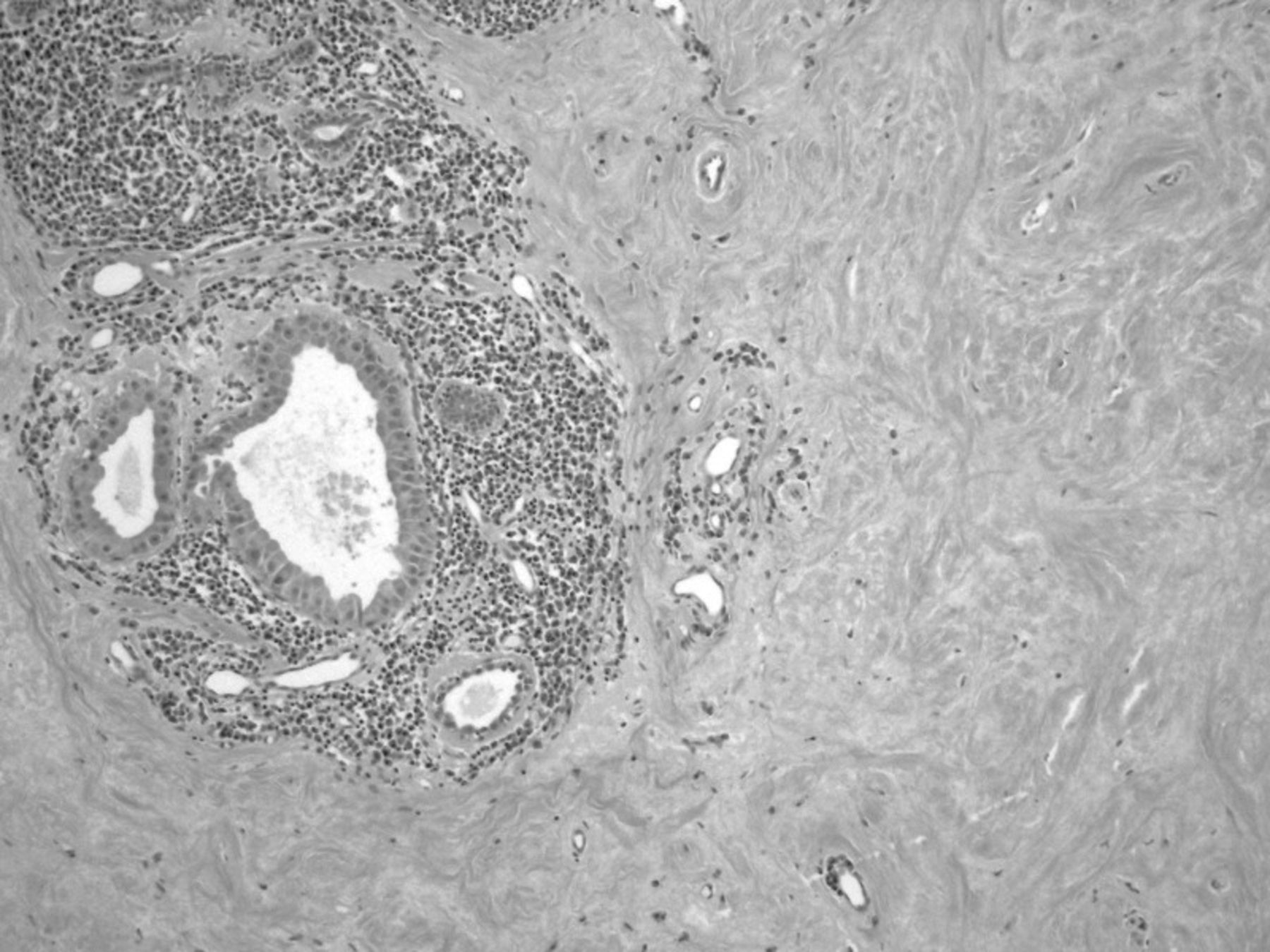
Standard hematoxylin and eosin histologic section of the lesion showing under low power the amyloid material in the breast, apocrine metaplasia, and periductal lymphoplasmacytic infiltration.

DISCUSSION

The deposition of insoluble protein characterized by systemic amyloidosis can interfere with physiological function. The clinical manifestations of amyloidosis are determined by the location of deposition.7 Systemic amyloidosis occurs when a monoclonal population of plasma cells generates excess amyloidogenic immunoglobulin light chains. In 75% of cases, the fibrillar deposits of systemic amyloidosis are composed of monoclonal lambda light chain proteins.7,8 In the remaining cases, protein deposits are products of kappa-restricted plasma cell clones. Kappa light chains have been described more frequently than lambda light chains with respect to amyloidosis of the breast.2 The incidence of systemic amyloidosis is 4.5 per 100,000, or approximately one-tenth the incidence of multiple myeloma.7

Primary breast amyloidosis is rare; however, it will typically present in women between the ages of 43 to 86 years old. Recent case reports have described instances of amyloidosis coexisting with breast cancer.2,4,9,10 Patients with breast amyloidosis are typically without clinical symptoms, with the first findings often noted on mammography or ultrasound. A minority of patients may present with a painless palpable mass or possibly a localized area of skin thickening, edema, or erythema.2 Primary amyloidosis of the breast is most often unilateral, but bilateral involvement has been reported.2,11-16

Although we describe a case of primary amyloidosis of the breast, the condition is most commonly associated with systemic amyloid involvement, manifesting outside of the breast in other parts of the body.2,11-16 Others have described amyloidosis as distinct lesions or occasionally as intermixed with breast cancer, and several different histologies have been reported, such as tubular carcinoma, invasive ductal carcinoma with extensive intraductal components, and invasive lobular carcinoma.10 Additional reports have included pathologies such as localized breast amyloidosis secondary to underlying extranodal Castleman disease, osseous metaplasia, and giant cell reaction.17,18 Other case reports have described isolated amyloidosis, or amyloidomas, of the lungs, larynx, urinary bladder, and heart.19-22

A recent review of 40 cases of breast amyloidosis over a 16-year period at the Mayo Clinic demonstrated an association with systemic amyloidosis in nearly 50% of all patients.5 Hematologic malignancies were also found in association with breast amyloidosis in a similar percentage of patients.5 As part of the systemic form, breast amyloid is frequently found as a late presentation. A case series by Charlot et al found that localized amyloidosis of the breast represented 0.5% of all the patients referred to the amyloid treatment center.23

CONCLUSION

The main treatment for primary amyloidosis of the breast is surgical removal, most often accomplished with breast conservation via lumpectomy. No role exists for associated examination of the draining nodal basin unless the final pathology reveals a concomitant breast cancer. In performing the lumpectomy, the goal should be to obtain negative surgical margins; however, no strong data exist to suggest that such lesions would recur with a microscopically positive margin. In this case, we were able to achieve negative margins. Adjuvant radiation therapy appears to be unnecessary. Postoperative management consists of periodic physical examination with a unilateral mammogram at 6 months to establish a baseline study. If this mammogram reveals postsurgical change and scar tissue, the patient can safely return to yearly bilateral screening mammograms.

This article meets the Accreditation Council for Graduate Medical Education and the American Board of Medical Specialties Maintenance of Certification competencies for Patient Care and Medical Knowledge.

Footnotes

  • The authors have no financial or proprietary interest in the subject matter of this article.

  • © Academic Division of Ochsner Clinic Foundation

REFERENCES

  1. ↵
    1. Fernandez BB,
    2. Hernandez FJ
    (2, 1973) Amyloid tumor of the breast. Arch Pathol 95(2):102–105, pmid:4683157.
    OpenUrlPubMed
  2. ↵
    1. Röcken C,
    2. Kronsbein H,
    3. Sletten K,
    4. Roessner A,
    5. Bässler R
    (5, 2002) Amyloidosis of the breast. Virchows Arch 440(5):527–535, pmid:12021928, Epub 2001 Oct 24.
    OpenUrlCrossRefPubMed
  3. ↵
    1. Sabaté JM,
    2. Clotet M,
    3. Gómez A,
    4. De Las Heras P,
    5. Torrubia S,
    6. Salinas T
    (Mar-Apr 2005) Radiologic evaluation of uncommon inflammatory and reactive breast disorders. Radiographics 25(2):411–424, pmid:15798059.
    OpenUrlCrossRefPubMed
  4. ↵
    1. Fu K,
    2. Bassett LW
    (10, 2001) Mammographic findings of diffuse amyloidosis and carcinoma of the breast. AJR Am J Roentgenol 177(4):901–902, pmid:11566701.
    OpenUrlPubMed
  5. ↵
    1. Said SM,
    2. Reynolds C,
    3. Jimenez RE,
    4. et al.
    (2, 2013) Amyloidosis of the breast: predominantly AL type and over half have concurrent breast hematologic disorders. Mod Pathol 26(2):232–238, pmid:23018872, Epub 2012 Sep 28.
    OpenUrlCrossRefPubMed
  6. ↵
    1. Lui PC,
    2. Pang LM,
    3. Hlaing TT,
    4. Tse GM
    (3, 2004) Tumorous amyloidosis of the breast associated with disseminated malignant lymphoma. J Clin Pathol 57(3):334–335, pmid:14990614.
    OpenUrlFREE Full Text
  7. ↵
    1. Stone MJ
    (2 1, 1990) Amyloidosis: a final common pathway for protein deposition in tissues. Blood 75(3):531–545, pmid:2137018.
    OpenUrlFREE Full Text
  8. ↵
    1. Merlini G,
    2. Stone MJ
    (10 15, 2006) Dangerous small B-cell clones. Blood 108(8):2520–2530, pmid:16794250, Epub 2006 Jun 22.
    OpenUrlAbstract/FREE Full Text
  9. ↵
    1. Munson-Bernardi BD,
    2. DePersia LA
    (1, 2006) Amyloidosis of the breast coexisting with ductal carcinoma in situ. AJR Am J Roentgenol 186(1):54–55, pmid:16357378.
    OpenUrlCrossRefPubMed
  10. ↵
    1. Sabate JM,
    2. Clotet M,
    3. Torrubia S,
    4. et al.
    (10, 2008) Localized amyloidosis of the breast associated with invasive lobular carcinoma. Br J Radiol 81(970):e252–e254, pmid:18796554.
    OpenUrlAbstract/FREE Full Text
  11. ↵
    1. Lew W,
    2. Seymour AE
    (Jan-Feb 1985) Primary amyloid tumor of the breast. Case report and literature review. Acta Cytol 29(1):7–11, pmid:3855589.
    OpenUrlPubMed
    1. McMahon RF,
    2. Connolly CE
    (6, 1987) Amyloid breast tumor. Am J Surg Pathol 11(6):488, pmid:3592063.
    OpenUrlPubMed
    1. McMahon RF,
    2. Waldron D,
    3. Given HF,
    4. Connolly CE
    (9, 1984) Localised amyloid tumour of breast—a case report. Ir J Med Sci 153(9):323–324, pmid:6490354.
    OpenUrlPubMed
    1. Silverman JF,
    2. Dabbs DJ,
    3. Norris HT,
    4. Pories WJ,
    5. Legier J,
    6. Kay S
    (8, 1986) Localized primary (AL) amyloid tumor of the breast. Cytologic, histologic, immunocytochemical and ultrastructural observations. Am J Surg Pathol 10(8):539–545, pmid:3526931.
    OpenUrlCrossRefPubMed
    1. Toohey JM,
    2. Ismail K,
    3. Lonergan D,
    4. Lewis CR
    (12, 2007) Amyloidosis of the breast mimicking recurrence in a previously treated early breast cancer. Australas Radiol 51(6):594–596, pmid:17958699.
    OpenUrlPubMed
  12. ↵
    1. Tutar E,
    2. Onat AM,
    3. Aydin A,
    4. et al.
    (2, 2008) Amyloid tumor of the breast mimicking breast carcinoma. South Med J 101(2):199–201, pmid:18364625.
    OpenUrlPubMed
  13. ↵
    1. Chiang D,
    2. Lee M,
    3. Germaine P,
    4. Liao L
    (2013) Amyloidosis of the breast with multicentric DCIS and pleomorphic invasive lobular carcinoma in a patient with underlying extranodal Castleman's disease. Case Rep Radiol 2013:190856. Epub 2013 Feb 4.
  14. ↵
    1. Deolekar MV,
    2. Larsen J,
    3. Morris JA
    (8, 2002) Primary amyloid tumour of the breast: a case report. J Clin Pathol 55(8):634–635, pmid:12147664.
    OpenUrlAbstract/FREE Full Text
  15. ↵
    1. Xu L,
    2. Frazier A,
    3. Burke A
    (1 15, 2013) Isolated pulmonary amyloidomas: report of 3 cases with histologic and imaging findings. Pathol Res Pract 209(1):62–66, pmid:23218762, Epub 2012 Dec 6.
    OpenUrlPubMed
    1. Park JH,
    2. Cheng HM
    (2013) Isolated laryngeal amyloidosis in a patient with a hoarse voice. BMJ Case Rep, Apr 5;2013.
    1. Toll AD,
    2. Ali SZ
    (2013) Urinary cytopathology in primary bladder amyloidosis. Acta Cytol 57(3):271–275, pmid:23636123, Epub 2013 Apr 25.
    OpenUrlPubMed
  16. ↵
    1. Podduturi V,
    2. Armstrong DR,
    3. Hitchcock MA,
    4. Roberts WC,
    5. Guileyardo JM
    (10, 2013) Isolated atrial amyloidosis and the importance of molecular classification. Proc (Bayl Univ Med Cent) 26(4):387–389, pmid:24082415.
    OpenUrlPubMed
  17. ↵
    1. Charlot M,
    2. Seldin DC,
    3. O'hara C,
    4. Skinner M,
    5. Sanchorawala V
    (6, 2011) Localized amyloidosis of the breast: a case series. Amyloid 18(2):72–75, pmid:21501022, Epub 2011 Apr 18.
    OpenUrlCrossRefPubMed
View Abstract
PreviousNext
Back to top

In this issue

Ochsner Journal
Vol. 14, Issue 2
Jun 2014
  • Table of Contents
  • Index by author
Print
Download PDF
Email Article

Thank you for your interest in spreading the word on Ochsner Journal.

NOTE: We only request your email address so that the person you are recommending the page to knows that you wanted them to see it, and that it is not junk mail. We do not capture any email address.

Enter multiple addresses on separate lines or separate them with commas.
Primary Amyloidosis of the Breast Presenting as a Solitary Nodule: Case Report and Review of the Literature
(Your Name) has sent you a message from Ochsner Journal
(Your Name) thought you would like to see the Ochsner Journal web site.
CAPTCHA
This question is for testing whether or not you are a human visitor and to prevent automated spam submissions.
Citation Tools
Primary Amyloidosis of the Breast Presenting as a Solitary Nodule: Case Report and Review of the Literature
Mary E. Huerter, Rasheed Hammadeh, Quan Zhou, Asher Weisberg, Adam I. Riker
Ochsner Journal Jun 2014, 14 (2) 282-286;

Citation Manager Formats

  • BibTeX
  • Bookends
  • EasyBib
  • EndNote (tagged)
  • EndNote 8 (xml)
  • Medlars
  • Mendeley
  • Papers
  • RefWorks Tagged
  • Ref Manager
  • RIS
  • Zotero
Share
Primary Amyloidosis of the Breast Presenting as a Solitary Nodule: Case Report and Review of the Literature
Mary E. Huerter, Rasheed Hammadeh, Quan Zhou, Asher Weisberg, Adam I. Riker
Ochsner Journal Jun 2014, 14 (2) 282-286;
del.icio.us logo Twitter logo Facebook logo Mendeley logo
  • Tweet Widget
  • Facebook Like
  • Google Plus One

Jump to section

  • Article
    • Abstract
    • INTRODUCTION
    • CASE REPORT
    • DISCUSSION
    • CONCLUSION
    • Footnotes
    • REFERENCES
  • Figures & Data
  • References
  • Info & Metrics
  • PDF

Cited By...

  • No citing articles found.
  • Google Scholar

More in this TOC Section

  • Propofol Infusion Syndrome or Not? A Case Report
  • Recurrent Angiomatoid Fibrous Histiocytoma: A Case Report and Review of the Literature
  • Late Recurrence of Light Chain Deposition Disease after Kidney Transplantation Treated with Bortezomib: A Case Report
Show more Case Reports

Similar Articles

Keywords

  • Amyloidosis
  • Congo red
  • mammogram
  • mastectomy–segmental

Our Content

  • Home
  • Current Issue
  • Ahead of Print
  • Archive
  • Featured Contributors
  • Ochsner Journal Blog
  • Archive at PubMed Central

Information & Forms

  • Instructions for Authors
  • Instructions for Reviewers
  • Submission Checklist
  • FAQ
  • License for Publishing-Author Attestation
  • Patient Consent Form
  • Submit a Manuscript

Services & Contacts

  • Permissions
  • Sign up for our electronic table of contents
  • Feedback Form
  • Contact Us

About Us

  • Editorial Board
  • About the Ochsner Journal
  • Ochsner Health
  • University of Queensland-Ochsner Clinical School
  • Alliance of Independent Academic Medical Centers

© 2025 Ochsner Clinic Foundation

Powered by HighWire