
Abstract
Background Foreign body aspiration (FBA) is a potentially life-threatening condition in children, and removal of an aspirated bead can be difficult.
Case Report An 11-month-old male infant presented with a history of choking 6 days prior to admission. FBA was suspected, and initial examination revealed a bead occluding the left main bronchus. The surgeon tried to retrieve the foreign body, but the patient developed coughing episodes with desaturation. The patient was intubated and a bronchoscopy was performed with a flexible bronchoscope. A Fogarty catheter was passed through the bronchoscope and then advanced through the bead opening. The distal balloon was inflated, and the bead was removed as the Fogarty catheter was withdrawn.
Conclusion We successfully removed an aspirated bead from an infant using the passing-through technique with a Fogarty catheter. Maintaining spontaneous ventilation for as long as possible and good coordination between the anesthesiologist and surgeon are crucial in such cases.
INTRODUCTION
Foreign body aspiration (FBA) is a potentially life-threatening event in children.1 FBA is a common cause of mortality and morbidity in children, especially in those younger than 2 years. Prompt diagnosis and early treatment are essential to minimize potentially serious consequences.2 Endobronchial foreign bodies must be secured and controlled during removal to avoid converting a partial airway obstruction into a complete airway obstruction. However, spherical objects may be difficult to retrieve because attempts to grasp the object may push it deeper into the bronchus. Fogarty catheters (Edwards Lifesciences) have been used in these situations.2
Good and Deutsch reported the case of a 7-year-old patient with FBA in whom controlled removal was accomplished by securing the bead between the balloon and the bronchoscope with the catheter tubing threaded through the lumen of the bead.2 We report the use of this technique to remove a bead from an 11-month-old patient.
CASE REPORT
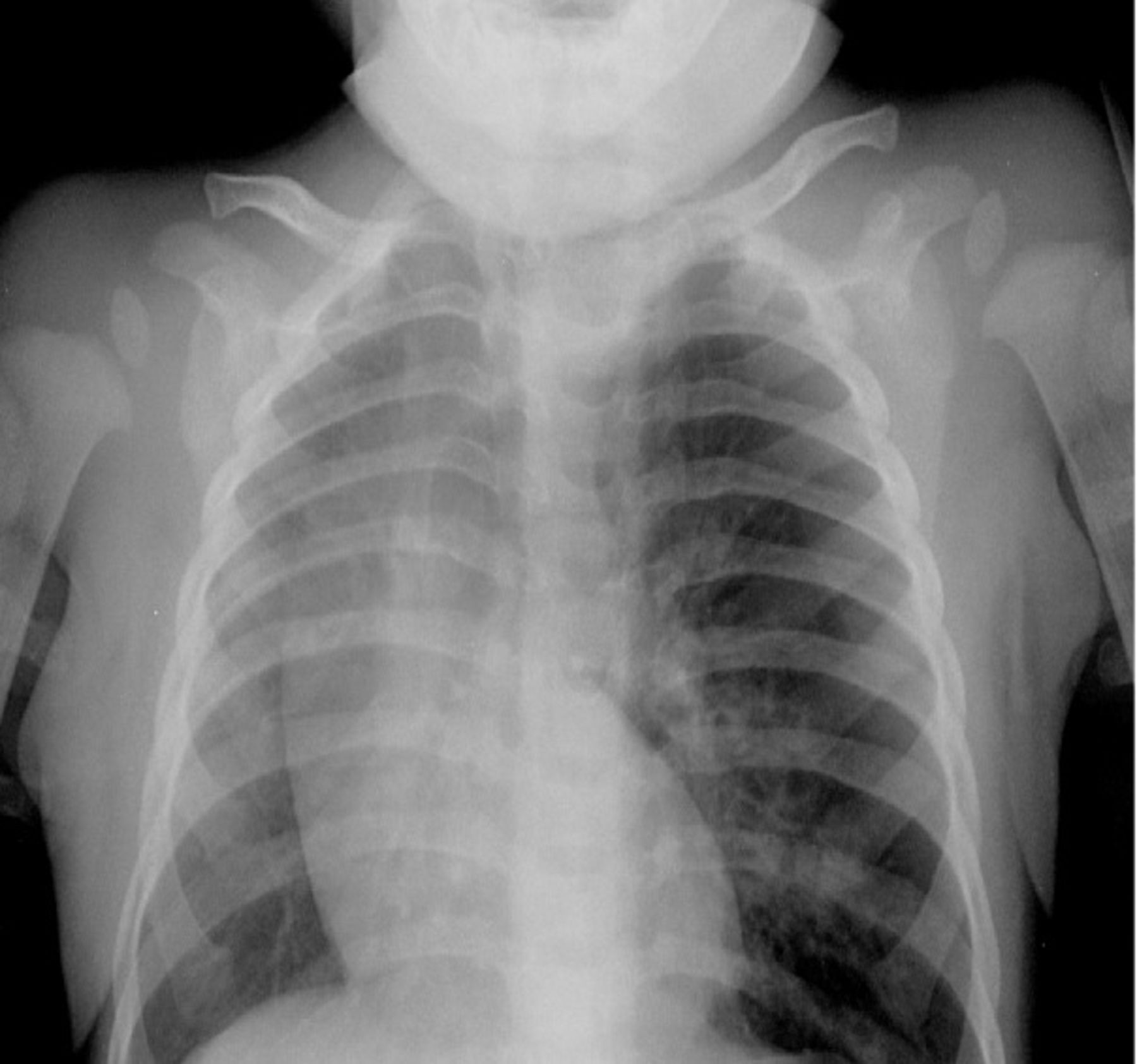
An otherwise healthy 11-month-old male infant, weighing 11 kg with negative birth, medical, and surgical history, had a choking/coughing episode 6 days prior to presentation that resolved when the patient's father performed the Heimlich maneuver. The patient was later taken to an outside hospital where a chest x-ray proved negative. He continued to have a hoarse cough and difficulty breathing. One day prior to this presentation, the patient had a low-grade fever with decreased oral intake. Chest x-ray at that time was consistent with left upper lobe hyperinflation. Although no obvious foreign body was identified on the x-ray, we thought the hyperinflation was caused by the ball-valve effect of a foreign body in the bronchus (Figure). The chest x-ray also showed a rightward shift of the heart, with a diagnostic impression of FBA. The patient was transferred to our institution for further management and scheduled for a direct laryngoscopy and bronchoscopy under general anesthesia.

Chest x-ray shows hyperinflation of left upper lobe, pushing the heart toward the right.
Premedication was given with intravenous (IV) midazolam 0.25 mg. Intraoperative management included standard American Society of Anesthesiologists monitoring. A gradual inhalation induction was performed to maintain spontaneous ventilation and allow the surgeon to visualize the foreign body without changing its position. Anesthesia was maintained with a propofol infusion of 350-400 μg/kg/min. The surgeon began with a suspension laryngoscopy, sprayed lidocaine, passed the vocal cords, and then completed a rigid bronchoscopy to identify the foreign body. The initial examination revealed a bead occluding the left main bronchus. The surgeon tried to retrieve the foreign body, but the patient developed episodes of breath-holding and coughing.
Despite increasing the propofol infusion, the patient's oxygen saturation started to drop into the 80s. We decided to give a muscle relaxant (rocuronium) and start jet ventilation at an initial pressure of 10 psi. This pressure was not enough to generate a good chest rise; however, oxygen saturation rose into the 90s. We decided to increase the psi to 15 with a respiratory rate of 30-40 jets per minute, allowing adequate time for exhalation. We monitored chest rise, oxygen saturation, and pericardial stethoscope. We were able to maintain oxygen saturation between 94%-95%. While the surgeon was preparing the Fogarty catheter and the flexible bronchoscope, we intubated the patient's trachea and used conventional ventilation. His oxygen saturation was brought to 99%-100%.
Flexible bronchoscopy was performed through the endotracheal tube to retrieve the foreign body. A 3-French, 12 in Fogarty catheter was advanced over the bead, and we attempted retrieval by inflating the distal balloon with 1 mL of normal saline. During manipulation, the bead spun, revealing a hole through it. The Fogarty catheter was then deflated and advanced through the bead's opening. The distal balloon was inflated again with 1 mL of normal saline, and the bead was removed as the Fogarty catheter was withdrawn. After the surgeon retrieved the foreign body, the muscle relaxant was reversed. The airway was suctioned, and the patient was extubated fully awake in the operating room. He was then transferred to the pediatric intensive care unit for observation. He remained in stable condition and was discharged home the next day.
DISCUSSION
FBA or foreign body ingestion is a common cause of morbidity and mortality in children, responsible for more than 17,000 emergency department visits per year of children younger than 14 years in the United States.3 Medical history is the single most predictive factor in clinical suspicions of FBA. Potential complications of FBA include pneumomediastinum, pneumothorax, total atelectasis, foreign body dislodgement, bronchiectasis, recurrent and/or unresolving pneumonia, and destruction of bronchial cartilage. If clinical history is suggestive of tracheobronchial foreign body aspiration, even in the presence of a negative physical examination and radiographic imaging, bronchoscopic evaluation is indicated.2
Bronchoscopic extraction of airway foreign bodies can be safely accomplished with both the rigid and flexible bronchoscope in adults and children. Rigid bronchoscopy has been successfully used since the early 1970s and remains the gold standard for foreign body removal.2 Rigid bronchoscopy allows for control of the airway and provides excellent visualization with a variety of available ancillary instruments. In addition, because of advances in fiber-optic techniques and improved instrumentation, bronchoscopy is a relatively safe procedure.4 However, even with improved bronchoscopic techniques, extraction is sometimes difficult because of fragmentation and migration of the foreign object. The Fogarty balloon-tipped catheter has been used for the retrieval of foreign bodies from many parts of the body.5
Reported complications in the use of Fogarty catheters for endobronchial foreign body removal include catheter disruption and tip embolization,6 excessive withdrawal force, and damage to the catheter from repeated sweeps of the dilated balloon.
Emergency bronchoscopy is necessary if the child is in acute respiratory distress. Otherwise, time should be allowed for fasting and rehydration in preparation for general anesthesia. Cooperation and communication between the surgeon and anesthesiologist are essential in FBA cases because the anesthesiologist and the surgeon are competing for the same airway.
Induction of anesthesia by inhalation or IV route is described in the literature. A survey of members of the Society for Pediatric Anesthesia found that most anesthesiologists prefer mask induction without cricoid pressure for a child with an aspirated foreign body.7 Keeping the patient spontaneously breathing while the suspension laryngoscope is introduced and the airway is manipulated is challenging. When intraoperative muscle paralysis medication is administered, ventilation can be controlled in a number of ways, including use of the microlaryngeal tube, insufflation of high flows of oxygen through a small catheter placed in the trachea, the intermittent apnea technique, manual jet ventilation,8 or extracorporeal membrane oxygenation when complete tracheal occlusion exists and other methods are ineffective.
These endoscopic procedures can be relatively long and may require a change of plan and equipment, as in our case. Therefore, anesthesiologists must take advantage of any opportunity when the surgeon is not working on the airway (eg, setting the equipment) to intubate the patient's trachea and conventionally ventilate the lungs to recruit the atelectatic alveoli. These steps are especially important in children because of their low functional residual capacity and high closing volume, both of which predispose them to rapid development of atelectasis and hypoxia.
Jet ventilation has been reported in adults but is not widely used in children, perhaps because of concerns that jet ventilation is likely to dislodge the foreign body; damage the tracheal mucosa; or cause subcutaneous emphysema, pneumomediastinum, or pneumothorax.9
No outcome data exist to support a decision between spontaneous or positive pressure ventilation; each has advantages and disadvantages. Inglis and Wagner10 reported that 98% of the bronchoscopies in their study were performed using spontaneous respiration, while only 2% required the use of myorelaxant drugs. In another study, muscle relaxants were used to treat more difficult cases.11 Soodan et al reported that anesthesia with mechanical ventilation might be more useful.12 All previously mentioned ventilation techniques were proven safe.
In our case, we kept the patient spontaneously breathing, maintained depth of anesthesia with local anesthesia of the airway, and gave a required muscle relaxant.
One of the complications of foreign body removal is obstruction of the airway caused by movement of the foreign body. Use of the Fogarty balloon in these cases appears to reduce the risks.
No strong evidence supports choosing one approach to general anesthesia over another for bronchoscopy of an inhaled foreign body. Careful preoperative planning and experience in pediatric airway management are crucial in preventing an adverse outcome and obtaining good results.
CONCLUSION
In this case, the chest x-ray showed symptoms suggestive of FBA without a foreign body visible on the image. It is very important to understand that foreign bodies may not show up on a chest x-ray. Maintaining spontaneous ventilation for as long as possible is advisable. Coordination between the surgeon and anesthesiologist is crucial for successful management of this type of case.
This article meets the Accreditation Council for Graduate Medical Education and the American Board of Medical Specialties Maintenance of Certification competencies for Patient Care and Medical Knowledge.
Footnotes
The authors have no financial or proprietary interest in the subject matter of this article.
- © Academic Division of Ochsner Clinic Foundation
In this issue


Jump to section
Cited By...
- No citing articles found.