
INTRODUCTION
Chiari II malformation is a complex constellation of findings resulting from an open neural tube defect that may manifest with malformations of the skull, dura, hindbrain, cerebellum, midbrain, posterior cerebrum, and spinal cord. An uncommonly imaged yet defining characteristic of the Chiari II malformation is a myelomeningocele.
HISTORY
A preterm, 31-week gestational age, infant male presented at birth with macrocephaly and a thoracic myelomeningocele. The patient was born to a 33-year-old gravida 3 para 3 mother who had received limited prenatal care. Physical examination demonstrated macrocephaly with bulging anterior fontanelle, scoliosis, adactyly with a single digit right foot, spontaneous bilateral upper extremity and lower extremity movements, and a thoracic myelomeningocele.
RADIOGRAPHIC APPEARANCE AND TREATMENT
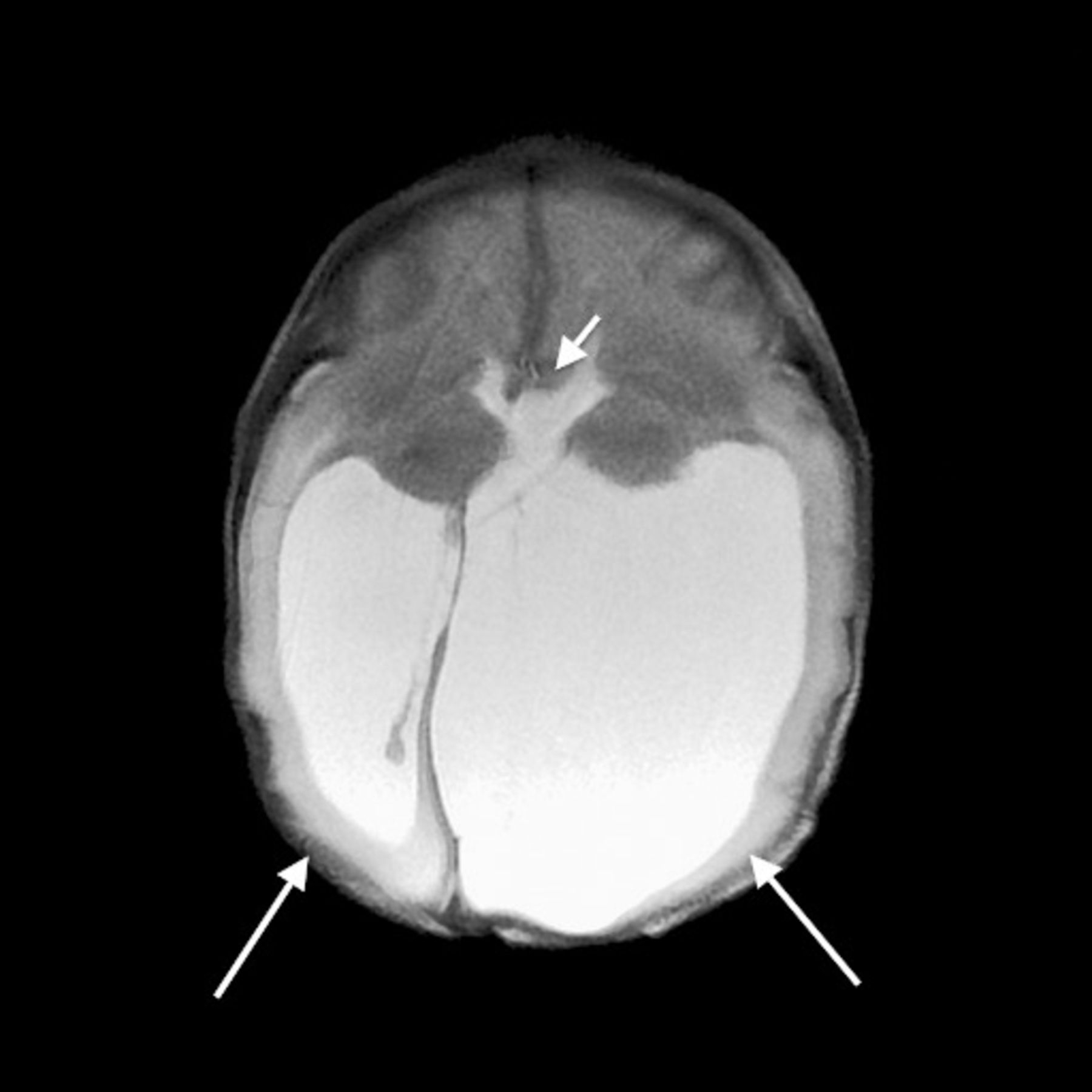
Given the patient's clinical appearance, magnetic resonance imaging (MRI) of the brain and spine was obtained shortly after delivery. MRI of the brain demonstrated marked ventriculomegaly with the choroid plexus floating in the enlarged lateral ventricles (Figure 1). The bilateral basal ganglia demonstrated increased separation, and the corpus callosum was not well established. Given this appearance, findings favored a diagnosis of callosal agenesis or callosal dysgenesis. Tectal beaking was also noted. A small posterior fossa was seen with widening of the foramen magnum and significant tonsillar herniation to the level of the C3 and C4 vertebral bodies (Figure 2), consistent with Chiari II malformation.

Axial T2 fast spin echo magnetic resonance image of the brain demonstrates extensive dilatation of the lateral (large arrows) and third ventricles (small arrow). The corpus callosum is not clearly established, suggesting callosal agenesis or callosal dysgenesis.

Sagittal T2 fast spin echo magnetic resonance image demonstrates a small posterior fossa with cerebellar tonsillar herniation extending through the foramen magnum to the level of the third cervical vertebral body (arrow).
MRI of the cervical, thoracic, and lumbar spine also demonstrated findings consistent with Chiari II malformation. Multiple vertebral body segmentation anomalies were seen, including a right T8 hemivertebra and a T5 butterfly vertebra. Marked dextroscoliotic curvature of the thoracolumbar spine was noted, most pronounced at the level of the T8 hemivertebra. A 1.2 cm posterior defect was seen originating at the level of T8. Posterior herniation of the spinal cord was noted at this level with extension beyond the soft tissues into a 3 × 2 cm cystic sac, compatible with a myelomeningocele (Figure 3A and 3B). The spinal cord reentered the spinal canal through the defect and terminated within the spinal canal at the level of T12. No evidence of diastematomyelia was seen.

Sagittal (view A) and axial (view B) T2 fast spin echo magnetic resonance images of the spine demonstrate spinal dysraphism with a posterior defect at the level of T8-T9 with dorsal herniation of the spinal cord through the posterior defect into a cystic space (arrow). The caudal portion of the cord is noted reentering the spinal canal and terminating at the level of T12 without evidence of diastematomyelia.
SURGERY
The patient was taken for myelomeningocele closure and shunt placement shortly after imaging. The two primary goals of surgery were to repair the spinal defects and exposed neural elements and to divert the cerebrospinal fluid (CSF) to treat the hydrocephalus. Although the repair of a thoracic-level defect such as in this patient is much more challenging than the usual lumbar/sacral myelomeningoceles, the same basic principles and steps apply: (1) dissection of the placode free from skin elements, (2) restoration of relatively normal spinal cord anatomy and position of the spinal canal, (3) a dura or dura-like closure, and (4) extensive soft tissue dissection to enable a primary skin closure. The hydrocephalus was addressed with a standard ventriculoperitoneal shunt placement immediately after the closure. The patient tolerated both procedures well without complication.
DISCUSSION
Chiari II is a complex malformation of the brain and spine secondary to an open neural tube defect. Myelomeningocele, the key component of Chiari II malformations, is seen in approximately 0.4 of every 1,000 live births, and associated hydrocephalus is noted in 85%-90% of patients.1 The findings manifest during the fourth week of fetal life because of failure of neural tube closure, resulting in CSF escape and insufficient cerebral ventricular expansion. A pressure gradient is created between intracranial and intraspinal spaces. In turn, reduced intraspinal CSF pressures lead to abnormal neural development.2
Cerebral manifestations include a small posterior fossa with parenchymal crowding that can lead to a widened foramen magnum and tonsillar herniation as seen in our patient.3 Tectal beaking is another distinguishing characteristic of Chiari II malformation3,4 and may relate to compression by the temporal lobes, as well as the rostral-caudal pressure gradient, that leads to stretching of the tectum in a posterior and inferior fashion.2 Additional supratentorial abnormalities are also commonly seen, including hypoplasia of the corpus callosum or hypogenesis that may be seen in 70%-90% of affected patients. A hypoplastic or fenestrated falx is another common finding, possibly secondary to chronic hydrocephalus and resulting in interdigitation of parasagittal cerebral gyri.2
Myelomeningoceles are seen in virtually all cases of Chiari II malformation,1 and improved surgical techniques have demonstrated improved outcomes in these patients. Radiologic evaluation plays a key role in the intrauterine diagnosis of Chiari II malformation, which may lead to intrauterine repair.5 Intrauterine diagnosis of myelomeningocele formation can help stratify patients based on myelomeningocele vertebral level, and stratification can help predict clinical outcomes such as leg function and shunt-dependent hydrocephalus.6 Patients with inferior myelomeningocele levels demonstrate lower and delayed incidences of shunt-dependent hydrocephalus.7 While MRI and ultrasound have demonstrated similar accuracy in determining myelomeningocele vertebral level, MRI has been shown to provide additional detail about intracranial abnormalities that may assist the family's decision to proceed with intrauterine repair.6
Although many myelomeningoceles are now diagnosed and treated in utero, our case presents a rare postgestational look at this distinguishing characteristic of the Chiari II malformation. Our case also represents a rare case of thoracic-level spinal dysraphism. In this case, the spinal cord demonstrates dorsal herniation through the spinal cord defect.
After postnatal closure of the myelomeningocele, >80% of patients develop hydrocephalus within the first 48 hours.5 Thus, radiologic surveillance becomes a key diagnostic and surveillance modality following myelomeningocele repair.
CONCLUSION
Better understanding of the clinical and radiographic nuances of spina bifida and Chiari II malformation is important to surgeons and radiologists who treat these complicated patients. Proper radiographic evaluation improves operative planning and follow-up surveillance of these patients.
- © Academic Division of Ochsner Clinic Foundation


