
Abstract
Background Vascular malformations are generally detected in childhood or adolescence with first presentations in adulthood being rare.
Case Report We report the case of a 52-year-old female with threatened compromise of her airway after expectorating a massive arteriovenous malformation anchored at the supraglottis. The only preceding symptom was dysphagia. The lesion was resected, the patient had a quick recovery, and she has shown no evidence of recurrence.
Conclusion Although uncommon, vascular malformations of the supraglottis or hypopharynx should be considered in the differential diagnosis of a patient presenting with dysphagia because of the potential to cause disastrous airway compromise. Although a lesion presenting acutely mandates a definitive airway plan, when clinically possible, computed tomography scan and indirect laryngoscopy can provide useful information for the airway and operative teams.
- Arteriovenous malformations
- deglutition disorders
- head and neck neoplasms
- obstructed airway
- vascular malformations
INTRODUCTION
Vascular malformations in the head and neck encompass a wide range of lesions and present an interesting challenge for the otolaryngologist. Such malformations can be formed from a single vessel type (arterial, venous, capillary, or lymphatic) or a combination and can occur anywhere within the body.1 Consequently, this varied group of lesions has the potential for numerous unique presentations with associated symptoms.
Capillary (infantile) hemangiomas and congenital hemangiomas classically undergo an initial proliferative phase followed by an involution phase that usually results in complete regression of the lesion. The natural history of other vascular malformations, however, can be volatile; some vascular malformations continue to proliferate without regression and often require intervention.2 Surgery is a widely accepted form of intervention;3 however, it has been correlated with high recurrence rates when performed as a primary procedure on lesions >5 cm.4 Other treatment options include embolization, sclerotherapy, observation, or a combination of these methods.5
Found in 1.5% of the population,6 vascular malformations are typically present at birth (90%).2 They are the result of altered proliferation, differentiation, maturation, and apoptosis of vascular cells1 and are recognized as benign lesions.2 Symptoms are varied and can include skin discoloration, bleeding, thrombophlebitis, or symptoms secondary to compression of surrounding structures.2 Vascular malformations have the potential to occur in any region of the body with predominance (60%) in the head and neck.5 Kobayashi and colleagues found in their population that most malformations arose in the oral cavity (59%) and nasal cavity (35%).4
CASE REPORT
A 52-year-old indigenous Australian female was brought to a regional hospital by ambulance after expectorating a large mass from her pharynx. Her history suggested the only preceding symptom was 6 months of dysphagia to solids for which she was about to undergo diagnostic testing by her family doctor. The initial assessment at the regional hospital was that the patient was stable with no hemorrhage or airway compromise from the mass, although the mass appeared to be anchored below the level of the tongue (Figure 1). The patient had expectorated the lesion and had to maintain traction on the mass to stop it from retracting into the pharynx and occluding her airway. She was transferred to a larger regional hospital unintubated and without airway complications.

The large expectorated lesion on presentation to the hospital.
On arrival at the treating hospital, a computed tomography (CT) scan (Figure 2) showed a lesion arising from the left anterior supraglottis. Flexible nasendoscopy revealed a mass arising from the left supraglottis; the glottis appeared uninvolved. Direct laryngoscopy and rapid-sequence intubation with a size 5 microlaryngoscopy endotracheal tube was successful on first attempt. The 18 cm fusiform lesion arose from a narrow base on the left aryepiglottic fold. Using suspension microlaryngoscopy technique, the operative team snared the lesion at its base with multiple Vicryl ties and resected it using monopolar electrocautery and cold steel through the operating laryngoscope. The patient lost <20 mL of blood during the entire procedure. She was extubated immediately following and suffered no postoperative complications. Histologically, the lesion consisted of fatty tissue with a predominance of vessel types, including thick- and thin-walled vessel types as well as capillaries. The lesion was most consistent with a nonhemangioma vascular malformation. The patient was discharged on postoperative day 2 and has since reported her dysphagia has completely resolved. She remained free of recurrence 9 months after the initial resection.
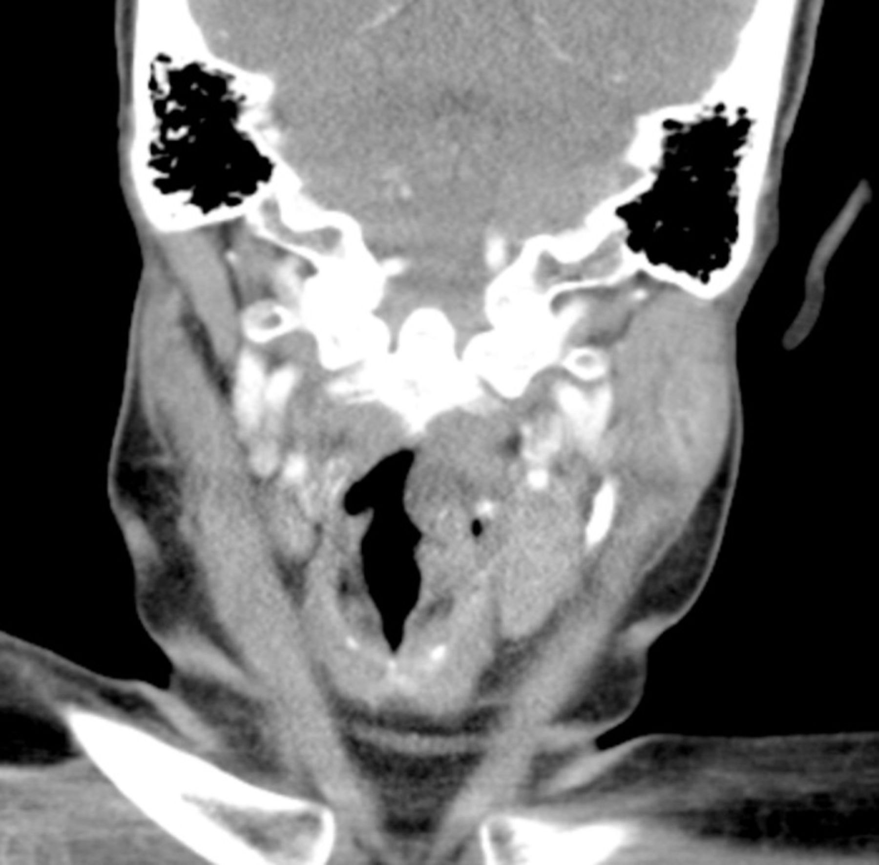
Computed tomography scan shows the lesion arising from the left anterior supraglottis.
DISCUSSION
Vascular malformations refer to a collection of abnormal vessels resulting from embryonic and fetal development defects.7 They are categorized according to vessel type: capillary, venous, arterial, lymphatic, or a combination.7 The International Society for the Study of Vascular Abnormalities divides these vessel abnormalities into vascular tumors and malformations. Malformations are further divided into slow-flow (capillary, venous, or lymphatic) or fast-flow (arterial) vessels; combinations of these are called arteriovenous malformations (AVMs), lymphaticovenous malformations, or capillary-lymphaticovenous malformations.8 Histologically, the lesion in this case was consistent with an AVM.
Vascular malformations typically exist at birth7 and usually present in childhood or adolescence. First presentation during adulthood is rare, especially with venous malformations in the pharyngolaryngeal region.9 Vascular malformations are occasionally associated with systemic disease or other anatomic abnormalities in syndromes such as Klippel-Trenaunay, Parkes Weber, Servelle-Martorell, and Sturge-Weber.8 Regression is not usually seen with lesions of this type, and they continue to proliferate. Although no treatment is required for these benign lesions, because of their size and the symptoms, many require definitive treatment.2
Symptoms of vascular malformations are predominantly caused by their compression of surrounding anatomy. In addition, each vessel type can produce its own symptoms.7 Venous malformations are considered the most common and are slow growing unless thrombosed.6,7 Largely, they are soft, compressible, and sometimes asymptomatic. Thrombophlebitis is a common complication of venous malformations in the head and neck and is associated with a distended, firm, and painful mass most prominent upon waking.7 Obstructive sleep apnea is another complication seen in upper aerodigestive tract venous malformations because they compress the upper airway.7 Arterial malformations are often detected by a palpable thrill, bruit, and warmth. Venous malformations also have the potential to enlarge rapidly after trauma or with hormonal changes, particularly during puberty. Severe pain and bleeding can be late features caused by ischemia and ulceration.7 Capillary malformations have few specific symptoms and will expand in a nodular pattern with discoloration as the most prominent sign. Vascular malformations can be associated with a range of symptoms, especially when comprised of multiple vessel types. Despite the size of her AVM, our patient's only reported symptom was a 6-month history of dysphagia.
Symptomatic lesions usually require intervention comprised of endovascular embolotherapy, surgery, sclerotherapy, or a combination.5 Low prevalence and institutional preference mean few randomized controlled trials support one intervention over another. Embolization is intended to halt growth and prevent hemorrhage. Zheng and colleagues suggest surgery should only be performed when embolization fails or endovascular access is limited, and they emphasize the difficulty associated with a vascular lesion that can involve various structures with poorly visible margins.5 They suggest surgery is indicated for well-defined lesions when anatomical structure can be largely preserved and when complete resection is the intent, because subsequent embolization is difficult if surgery fails. Embolization is commonly used in conjunction with operative management, especially when intraoperative or postoperative hemorrhage may be hard to control.2 Recurrence is common after surgery, as complete excision of the lesion is often difficult but is essential for complete resolution.5 Excision of the multiple feeder and draining vessels is not necessary to prevent recurrence; however, they can obstruct the surgical field and make resection difficult.5 Kobayashi and colleagues demonstrated in their population that only lesions <5 cm are likely to resolve after one treatment.4 Other studies have implied that surgical intervention resulting in complete resolution of AVMs is rare.2,5,10 The lesion presented in this case was well demarcated with little alteration of the surrounding anatomy. The AVM was primarily resected with negligible intraoperative hemorrhage and no subsequent recurrence at 9-month follow-up, making it a good example of when primary resection may be favored over endovascular therapies.
Securing the patient's airway is often problematic when dealing with lesions arising from the aerodigestive tract. McLoughlin and McBrien described a case with an orofacial AVM causing sudden-onset stridor and requiring supplemental oxygen following an acute ischemic infarct of the left frontal lobe.11 The AVM involved the patient's left face, neck, and tongue; however, the extent of this lesion could not be determined with CT prior to securing the airway. Because of the unknown size and location of the AVM, the surgeons were reluctant to perform a tracheostomy unless their first preference of endotracheal intubation failed. Although tracheostomy with local anesthesia is advised for obstructions arising from the glottis and supraglottis,1,12 our case demonstrated that the use of indirect laryngoscopy and CT scan can provide essential information for creating a definitive airway plan for patients whose airway is not immediately threatened. Rapid-sequence induction and endotracheal intubation proved to be the simplest and safest method to minimize complications in our patient's threatened airway and greatly reduced her postoperative morbidity when compared with a possible tracheostomy.
CONCLUSION
Although uncommon, vascular malformations of the supraglottis or hypopharynx should be considered in the differential diagnosis of a patient presenting with dysphagia because of the potential to cause disastrous airway compromise. For this supraglottic fusiform lesion, a primary resection resulted in a positive patient outcome and demonstrated that primary resection of AVMs should be considered when dealing with AVMs arising from the aerodigestive tract. Although a lesion presenting acutely such as this one mandates a definitive airway plan, when clinically possible, CT scan and indirect laryngoscopy can provide useful information for the airway and operative teams.
This article meets the Accreditation Council for Graduate Medical Education and the American Board of Medical Specialties Maintenance of Certification competencies for Patient Care and Medical Knowledge.
ACKNOWLEDGMENTS
The authors have no financial or proprietary interest in the subject matter of this article.
- © Academic Division of Ochsner Clinic Foundation
In this issue


Jump to section
Cited By...
- No citing articles found.