
Abstract
Background: Ameloblastic fibrosarcoma (AFS) is a rare odontogenic neoplasm of the jaw that usually arises de novo or through a malignant change in the mesenchymal component of a preexisting or recurrent benign fibroma. The majority of AFS cases reported in the literature arise in the mandible.
Case Report: A 35-year-old male presented with an asymptomatic left maxillary mass that on imaging was found to be effacing most of his maxillary sinus. He underwent a left maxillectomy with free-flap reconstruction and adjuvant radiotherapy to the tumor bed.
Conclusion: Wide local excision remains the treatment of choice for AFS, given the poor survival rates of patients with recurrent disease. However, long-term studies and follow-up are needed to elucidate the role of adjuvant therapies in the primary treatment of AFS.
INTRODUCTION
Ameloblastic fibrosarcoma (AFS) is a rare odontogenic neoplasm characterized by marked cytologic atypia, increased cellularity with diminution of the epithelial component, and aggressive behavior. Approximately 60 cases of AFS have been reported in the literature to date, with most cases located in the mandible.1 This low-grade sarcoma also has a slightly male preponderance with a mean age of 27.3 years.2 The main symptoms during presentation are pain, swelling, and rapid growth. If inadequately treated, AFSs show a high rate of local recurrence, but distant metastasis is extremely rare. The optimal treatment for patients with this type of sarcoma is usually wide surgical excision and long-term follow-up.3 The value of adjuvant treatment with chemotherapy or radiation therapy remains uncertain, and adjuvant therapy is generally only used in patients with multiple episodes of recurrence after initial resection.4
CASE REPORT
A 35-year-old male presented with an asymptomatic left maxillary mass extending into the sinus cavity. He had initially presented to his dentist with a swelling in the upper left buccal sulcus. Radiographic examination consisting of an orthopantomogram (OPG) showed bony destruction of the left maxilla (Figure 1). The OPG findings were initially thought to represent an abscess or cyst, and the patient was referred to an oral and maxillofacial surgeon for further management. A prebiopsy computed tomography (CT) scan showed an expansile lesion in the left maxilla effacing most of the maxillary sinus. The maxillofacial surgeon removed most of the mass and sent the tissue for histology. AFS in the left maxilla was diagnosed based on the histopathology. The histologic pattern of AFS superficially resembles ameloblastic fibroma with nests and cords of odontogenic epithelium set in a connective tissue background. The epithelial tissue was benign (Figure 2A), but the connective tissue component was malignant with hypercellularity of the mesenchymal components, some variability in nuclear size and shape, and obvious mitotic activity (Figure 2B). Chest CT and positron emission tomography scans showed no evidence of metastatic neoplastic disease.
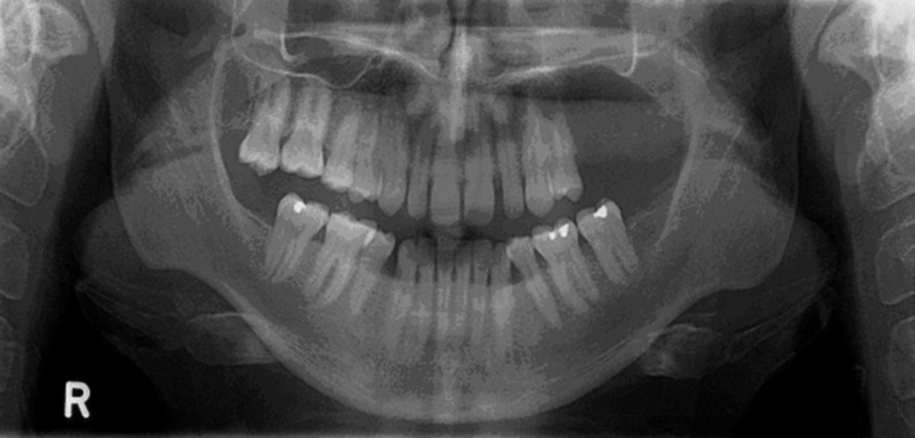
Preoperative orthopantomogram shows cortical thinning and bony destruction of the left side of the alveolar process of the maxilla.

A: Islands of ameloblastic epithelium set in a connective tissue background. The overall pattern is ameloblastic fibroma–like, but some of the background is more cellular than would normally be expected in a benign lesion. B: Small ameloblastic strands set in a markedly hypercellular background with some mitotically active cells.
The patient subsequently underwent a left maxillectomy with reconstruction with a left deep circumflex iliac artery bone free flap, using the iliac crest to reconstruct the parts of the maxilla that were resected. A composite free flap was chosen because of the extent of the resection that included the whole maxilla and the orbital floor on the left side. In addition, a bony reconstruction allows the future placement of dental implants. Histology of the resected maxilla showed a biphasic neoplasm with a prominent stromal component and a less prominent epithelial component. Whether the hypocellular areas without atypia or mitotic activity represented heterogeneity within the stroma or a preexisting benign lesion is uncertain. Although the tumor appeared to be completely removed, radiotherapy was recommended because of the proximity of the margin. Accordingly, 63 Gy in 35 fractions was administered to the tumor bed.
DISCUSSION
AFS is a rare odontogenic neoplasm derived from the ectomesenchyme of the dental pulp. In our case, the AFS arose within the maxilla, a considerably rare occurrence, as a majority of reported cases have been located in the molar and premolar regions of the mandible.3 While our case most likely arose de novo, a number of cases have been reported in which AFS arose from a preexisting ameloblastic fibroma.2
Radiologic features that are suggestive of possible lytic tumors are usually not diagnostic, as in our case, in which the initial findings on the OPG were thought to represent a cyst or abscess. The diagnosis was made from the histologic features that distinguish AFS from other sarcomas, fibromas, and blastomas of the maxillofacial region.
A wide surgical excision with clear margins seems to be the most reliable and significant prognostic factor for a favorable survival rate. Wide excision and clear margins are especially important because the survival rate in patients with local recurrence is poor.5
Adjuvant therapy in the treatment of AFS has generally been reserved for patients who have unresectable disease or need salvage operations following the initial surgery. High-dose radiation therapy to the tumor bed may help reduce the recurrence rate and local extension of the tumor.6 However, the decision to administer radiation therapy in a young patient is a balance between reducing the risk of local recurrence vs increasing the chances of a secondary malignancy years later.
To our knowledge, only 1 case of chemotherapy used to treat a patient with ameloblastic fibroma that underwent transformation to an ameloblastic fibrosarcoma during recurrence has been reported.4 Actinomycin D, vincristine, and cyclophosphamide were the chemotherapeutic agents used to produce a 50% reduction in the tumor mass. However, the clinical course was complicated by metastatic melanoma that resulted in the patient's death.
CONCLUSION
Definitive treatment of this disease remains difficult to define because of limited data and long-term follow-up of patients with this rare sarcomatous neoplasm. Surgical resection remains the standard treatment, as no clear or consistent pattern exists for the use of adjuvant therapy in the primary treatment of AFS. However, in this case, early and aggressive treatment of this cancer with both surgery and adjuvant radiotherapy was considered the best treatment modality. Our patient will need ongoing close and long-term follow-up. We hope this treatment will be sufficient to prevent recurrence and give our patient the best chance of long-term survival.
This article meets the Accreditation Council for Graduate Medical Education and the American Board of Medical Specialties Maintenance of Certification competencies for Patient Care and Medical Knowledge.
ACKNOWLEDGMENTS
The authors have no financial or proprietary interest in the subject matter of this article.
- © Academic Division of Ochsner Clinic Foundation


