
Main menu

  • Home
  • Content
    • Current
    • Ahead of print
    • Archive
  • Info for
    • Authors
    • Reviewers
  • About Us
    • About the Ochsner Journal
    • Editorial Board
  • More
    • Alerts
    • Feedback
  • Other Publications
    • Ochsner Journal Blog

User menu

  • My alerts
  • Log in

Search

  • Advanced search
Ochsner Journal
  • Other Publications
    • Ochsner Journal Blog
  • My alerts
  • Log in
Ochsner Journal

Advanced Search

  • Home
  • Content
    • Current
    • Ahead of print
    • Archive
  • Info for
    • Authors
    • Reviewers
  • About Us
    • About the Ochsner Journal
    • Editorial Board
  • More
    • Alerts
    • Feedback
Case ReportCASE REPORTS AND CLINICAL OBSERVATIONS

Xanthogranulomatous Osteomyelitis of the Tibia

Saeed Solooki, Amir Human Hoveidaei, Bahareh Kardeh, Negar Azarpira and Ehsan Salehi
Ochsner Journal September 2019, 19 (3) 276-281; DOI: https://doi.org/10.31486/toj.18.0165
Saeed Solooki
1Department of Orthopedic Surgery, Bone and Joint Diseases Research Center, Shiraz University of Medical Sciences, Shiraz, Iran
MD
  • Find this author on Google Scholar
  • Find this author on PubMed
  • Search for this author on this site
Amir Human Hoveidaei
2Student Research Committee, Shiraz University of Medical Sciences, Shiraz, Iran
3Students’ Scientific Research Center, Tehran University of Medical Sciences, Tehran, Iran
MD
  • Find this author on Google Scholar
  • Find this author on PubMed
  • Search for this author on this site
  • For correspondence: a.h.hoveidaei{at}sums.ac.ir
Bahareh Kardeh
4Bone and Joint Diseases Research Center, Clinical Neurology Research Center, Shiraz University of Medical Sciences, Shiraz, Iran
MD
  • Find this author on Google Scholar
  • Find this author on PubMed
  • Search for this author on this site
Negar Azarpira
5Transplant Research Center, Shiraz University of Medical Sciences, Shiraz, Iran
MD
  • Find this author on Google Scholar
  • Find this author on PubMed
  • Search for this author on this site
Ehsan Salehi
1Department of Orthopedic Surgery, Bone and Joint Diseases Research Center, Shiraz University of Medical Sciences, Shiraz, Iran
MD
  • Find this author on Google Scholar
  • Find this author on PubMed
  • Search for this author on this site
  • Article
  • Figures & Data
  • References
  • Info & Metrics
  • PDF
Loading

Abstract

Background: Xanthogranulomatous osteomyelitis (XO) is a rare chronic inflammatory process that is histologically characterized by the presence of foamy macrophages, histiocytes, and plasma cells. Radiologic and gross examinations can mimic malignancy, so definitive diagnosis should be made by histopathologic evaluation.

Case Report: A 15-year-old male presented with pain in the proximal right leg for 2 weeks prior to admission. The patient had a history of leg trauma 3 years prior that was responsive to as-needed analgesics. Laboratory data revealed increased erythrocyte sedimentation rate and C-reactive protein. X-ray of the right tibia showed a periosteal reaction and bulging of bone with a questionable destruction of the cortex. Magnetic resonance imaging demonstrated an expansile bony lesion in the proximal metaphysis of the tibia. Histopathologic examination showed the bone trabecula surrounded by chronic inflammatory cells and a fragment of dead bone surrounded by histiocytes, foamy macrophages, and plasma cells. The pathologist confirmed the diagnosis of XO. Wound culture proved moderate growth of Staphylococcus aureus, and treatment with cefazolin and cephalexin was successful.

Conclusion: The patient was not treated with the routine therapy used in previously reported cases (curettage), suggesting that antibiotic therapy should be considered before employing surgical interventions for XO.

Keywords:
  • Bone diseases
  • infection
  • osteomyelitis
  • tibia
  • xanthomatosis

INTRODUCTION

Xanthogranulomatous process is a rare form of chronic inflammation that is histologically characterized by the presence of foamy periodic acid-Schiff–positive histiocytes admixed with plasma cells and lymphocytes. This condition is predominantly observed in soft tissues and has been described in the gallbladder, kidney, lung, and gastrointestinal and urogenital tracts. Bone involvement is less common.1-4 Cozzutto initially coined the term xanthogranulomatous osteomyelitis (XO) in 1984.5 To the best of our knowledge, only 20 cases of XO have been reported since. We report a case of XO of the tibia in a young male.

CASE REPORT

A 15-year-old male with body weight of 65 kg and height of 160 cm was referred to our orthopedic center with the chief complaint of pain in the proximal right leg for the previous 2 weeks. On physical examination, the patient had tenderness, swelling, and erythema in the area. The patient reported a history of direct trauma to his right leg 3 years prior during a football game by one of the opposing team's players. The trauma was not severe and responded well to as-need oral analgesics, so the patient had not had the injury evaluated by a physician.

Laboratory investigation showed an elevated erythrocyte sedimentation rate (ESR) of 57 mm/hr (normal range, 0-20 mm/hr) and a C-reactive protein (CRP) level of 105 mg/L (normal range, ≤12 mg/L). Total leukocyte count and differential were within normal ranges; however, mild anemia was detected (red blood cells of 5.14 million/L, hemoglobin of 12.3 g/dL, and mean corpuscular volume of 74.9 fL). Renal and liver function tests were normal. Chest x-ray was negative.

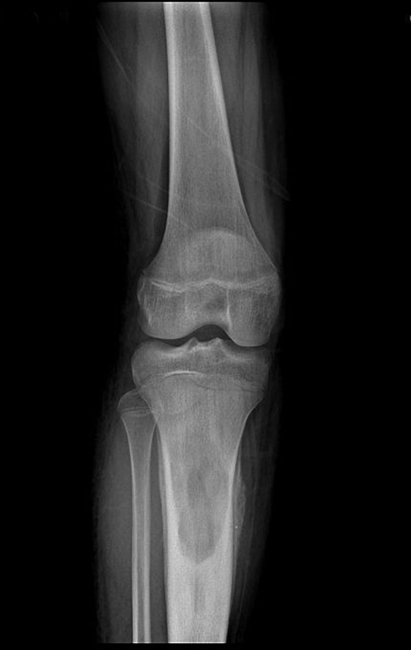
X-ray of the right tibia showed a periosteal reaction and bulging of the bone with a questionable destruction of the cortex (Figure 1). Magnetic resonance imaging revealed a 60 × 30 mm expansile bony lesion in the proximal metaphysis of the tibia, suggestive of osteomyelitis with abscess formation, Ewing sarcoma, or osteosarcoma (Figure 2). Bone scan exhibited increased uptake in the proximal right tibia.

Figure 1.
  • Download figure
  • Open in new tab
  • Download powerpoint
Figure 1.

X-ray on the day of admission showed periosteal reaction and bulging of the bone with a questionable destruction of the cortex.

Figure 2.
  • Download figure
  • Open in new tab
  • Download powerpoint
Figure 2.

Magnetic resonance imaging revealed an expansile bony lesion in the proximal metaphysis of the tibia.

The lesion was biopsied. Purulent discharge and multiple bony fragments with a creamy-gray color were grossly visible. Histopathologic examination showed the bone trabecula surrounded by chronic inflammatory cells and a fragment of dead bone surrounded by histiocytes, foamy macrophages, and plasma cells (Figures 3 and 4). The pathologist confirmed the diagnosis of XO.

Figure 3.
  • Download figure
  • Open in new tab
  • Download powerpoint
Figure 3.

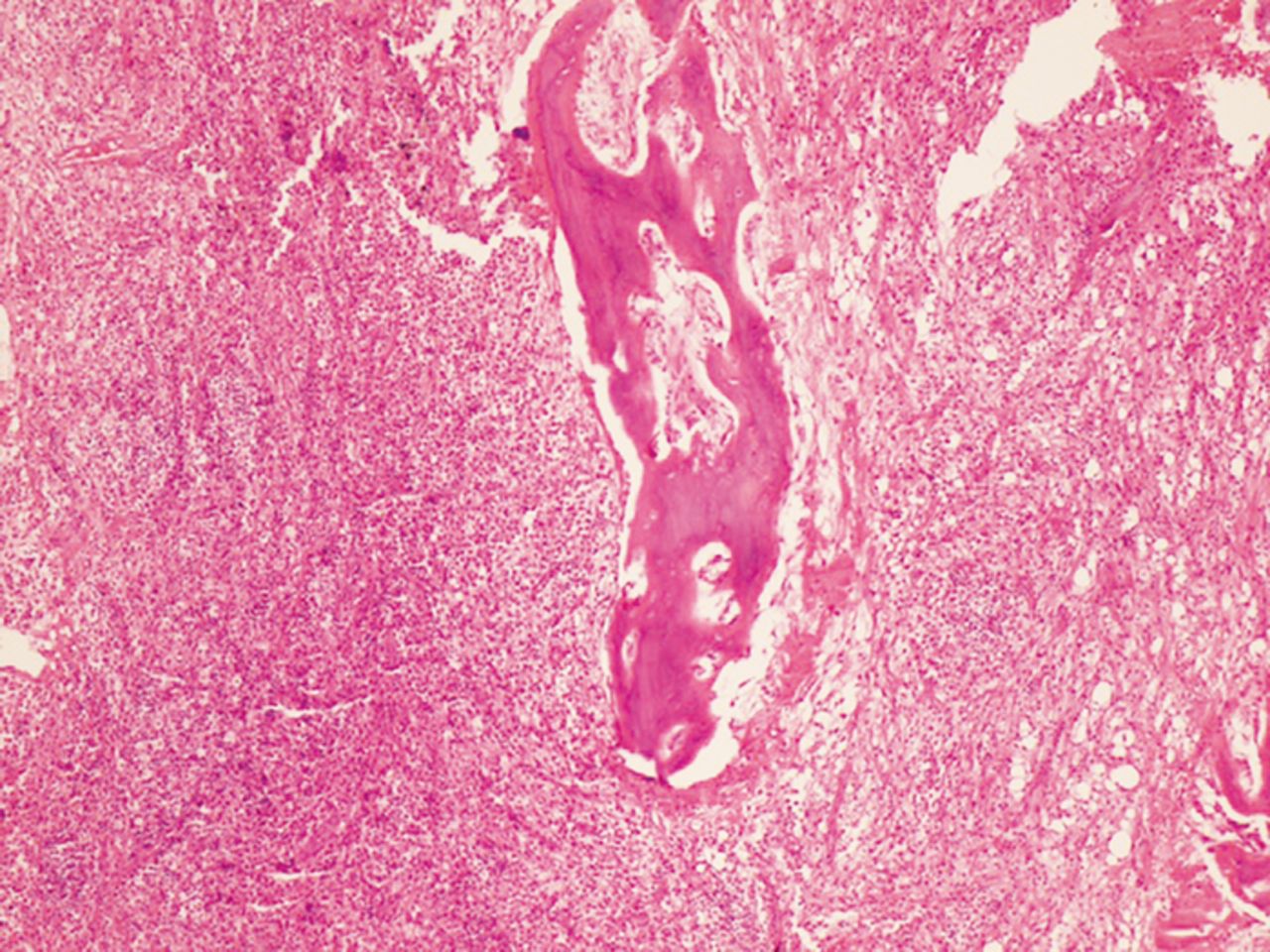
Lesion biopsy showed the bone trabecula surrounded by chronic inflammatory cells.

Figure 4.
  • Download figure
  • Open in new tab
  • Download powerpoint
Figure 4.

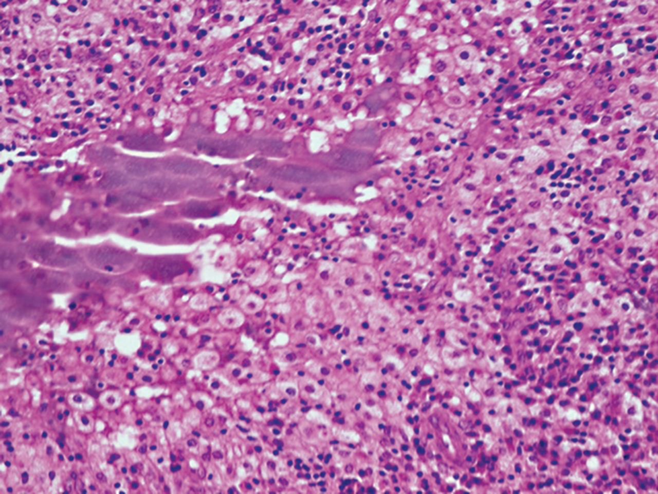
Lesion biopsy showed a fragment of dead bone surrounded by histiocytes, foamy macrophages, and plasma cells.

After biopsy demonstrated the infectious nature of the lesion, microbiologic assessment was requested, and the patient was administered intravenous (IV) cefazolin 500 mg every 8 hours and gentamicin 40 mg every 8 hours. Qualitative polymerase chain reaction was negative for tuberculosis. Wound culture proved moderate growth of Staphylococcus aureus that was sensitive to cefazolin and cephalexin. When these results were received after 12 days, gentamicin was changed to cephalexin 1 g IV every 8 hours, and cefazolin was continued at the same dosage and interval for 9 more days.

The patient's condition gradually improved during his hospital stay, and he was discharged on oral cephalexin and clindamycin for 3 weeks.

During the patient's 3-week admission, he received IV gentamicin for 12 days (which was not the proper choice according to the antibiogram), IV cefazolin for 21 days, IV cephalexin for 9 days, and oral cephalexin and clindamycin for 21 days.

At 11-week follow-up, the patient's symptoms had completely resolved. X-ray of the right tibia did not indicate any recurrence (Figure 5). In conjunction with the absence of symptoms, consecutive results of 0 mm/hr for ESR and 1 mg/L for CRP level at 11, 19, and 25 weeks of follow-up confirmed the success of the treatment.

Figure 5.
  • Download figure
  • Open in new tab
  • Download powerpoint
Figure 5.

Follow-up x-ray after 11 weeks showed no bone bulging or periosteal reaction.

DISCUSSION

XO was first introduced in the reports of 2 cases in 1984 followed by reports of 18 other patients summarized in the Table.5-22 A review of these 20 cases and the case of our patient suggests that XO is more prevalent in males (15 of 21) than females and has no characteristic age distribution. Most patients (17 of 21) had unifocal XO; only 4 had multifocal lesions.7,10,17,20 The most commonly affected sites are the long tubular bones (17 of 21). As with our patient, pain and swelling of the involved bone seem to be the primary reason for presentation.5-22

View this table:
  • View inline
  • View popup
Table.

Cases of Xanthogranulomatous Osteomyelitis Reported in the Literature

Cheema et al attributed the pathogenesis of xanthogranulomatous process in bone to a delayed-type hypersensitivity reaction of cell-mediated immunity.22 Trauma has been also mentioned as a hypothetical reason for this process. In addition to our case, 2 other patients with XO had a history of trauma.6,10 Trauma also seems to play a role in the development of xanthogranulomatous process in organs; Franco et al reported the role of gallstone trauma in inducing xanthogranulomatous cholecystitis.23 In some cases of XO, the bacterial organisms Mycobacterium marinum,7 S aureus,9,11 and Pseudomonas15 have been detected in specimen cultures; in our case, the culture was positive for S aureus. The association between bacterial infections and xanthogranulomatous lesions in the bone may become established as a probable etiopathogenesis, as with other organs.20

Given the possibility of multicentric lesions, whole-body radiologic analysis is recommended for a patient with confirmed XO; however, diagnosing XO with imaging is challenging.21 Radiologically and grossly, XO manifests as mass-like lesions mimicking infiltrative malignant tumors. Thus, differentiating this pseudoneoplasm of infectious etiology from true malignancy is a crucial step for implementing timely and appropriate management. Clinical background that includes history of trauma, radiographic evaluation, and histopathologic examination are essential diagnostic components.

Differential diagnoses of XO include the following:

  1. Langerhans cell histiocytosis (LCH): LCH lesions consist of Langerhans cells with eosinophil infiltration. Radiographically, LCH usually has circumscribed lytic bony lesions without surrounding sclerosis.24

  2. Xanthoma: Xanthoma can occur in the bone secondary to hyperlipidemia; however, xanthoma may not show suppurative inflammation.25

  3. Erdheim-Chester disease (ECD): ECD is a multifocal disorder with frequent involvement of extraskeletal tissues; histology shows foamy histiocytes, fibrosis without neutrophilic infiltrations, and cholesterol clefts.26

  4. Malakoplakia: In patients with malakoplakia, the histologic examination shows large foamy macrophages and Michaelis-Gutmann bodies.27

  5. Infiltrative lipid storage diseases: Clinical background (including muscle atrophy, cardiomyopathy, and dysfunction of internal organs), foamy macrophages of the bone marrow, and the absence of suppurative inflammation can help differentiate infiltrative lipid storage diseases from XO.28

  6. Metastatic renal cell carcinoma: Metastatic renal cell carcinoma can be easily differentiated from XO by history taking and histologic tumor patterns.29

  7. Chronic recurrent multifocal osteomyelitis (CRMO): CRMO is an autoinflammatory disease that usually occurs in a multicentric manner in the long bones and the shoulder girdle during childhood. The radiologic findings of CRMO are osteolytic initially but later demonstrate a hyperostotic and sclerotic state.30

  8. Fibrohistiocytic tumor: Fibrohistiocytic tumors are composed of cells that have morphologic features of fibro-blasts and histiocytes. However, these cells are not histio-cytes and may exhibit phagocytic properties, including neutrophil phagocytosis.4,31

Because of the rarity of XO, no uniform treatment protocol is available. Most cases report treatment with curettage and bone grafting. Antibiotics were prescribed in addition to surgical procedures (synovectomy and arthrodesis) for 2 patients.7,15 For 2 patients in addition to our case, treatment was completely based on antibiotics with regular follow-up.8,22

Although recommending the omission of surgical treatment is not possible, the authors do recommend requesting microbiologic assessment for all lesions with an infectious nature. Treatment with wide-spectrum antibiotics followed by treatment with definitive effective antibiotics is the best choice, followed by regular physical examination and laboratory follow-up, including ESR and CRP.

Based on 3 reported cases (including ours),8,22 a key point in the treatment of XO is to consider using antibiotic therapy as a noninvasive treatment before surgical intervention.

CONCLUSION

We report a case of XO because of its rarity, importance, and curability. While XO is benign, it can mimic malignant primary or secondary bone lesions in its clinical manifestations, gross examination, and imaging. Hence, clinicians and radiologists must be aware of XO and include it in their differential diagnosis.

This article meets the Accreditation Council for Graduate Medical Education and the American Board of Medical Specialties Maintenance of Certification competencies for Patient Care and Medical Knowledge.

Acknowledgments

The authors have no financial or proprietary interest in the subject matter of this article.

The authors would like to thank Shiraz University of Medical Sciences, Shiraz, Iran, the Center for Development of Clinical Research of Nemazee Hospital, and Dr Nasrin Shokrpour for editorial assistance.

  • © Academic Division of Ochsner Clinic Foundation

References

  1. 1.↵
    1. Ballentine WK 3rd.,
    2. Vilson F,
    3. Dyer RB,
    4. Mirzazadeh M
    . Nephron-sparing management of xanthogranulomatous pyelonephritis presenting as spontaneous renal hemorrhage: a case report and literature review. BMC Urol. 2018 Jun 5;18(1):57. doi: 10.1186/s12894-018-0354-3.
    OpenUrlCrossRef
  2. 2.
    1. Guzmán-Valdivia G
    . Xanthogranulomatous cholecystitis: 15 years’ experience. World J Surg. 2004 Mar;28(3):254-257. doi: 10.1007/s00268-003-7161-y.
    OpenUrlCrossRefPubMed
  3. 3.
    1. Bourm KS,
    2. Menias CO,
    3. Ali K,
    4. Alhalabi K,
    5. Elsayes KM
    . Spectrum of xanthogranulomatous processes in the abdomen and pelvis: a pictorial review of infectious, inflammatory, and proliferative responses. AJR Am J Roentgenol. 2017 Mar;208(3):475-484. doi: 10.2214/AJR.16.17075.
    OpenUrlCrossRef
  4. 4.↵
    1. Cozzutto C,
    2. Carbone A.
    The xanthogranulomatous process. Xanthogranulomatous inflammation. Pathol Res Pract. 1988 Aug;183(4):395-402. doi: 10.1016/S0344-0338(88)80085-2.
    OpenUrlCrossRefPubMed
  5. 5.↵
    1. Cozzutto C
    . Xanthogranulomatous osteomyelitis. Arch Pathol Lab Med. 1984 Dec;108(12):973-976.
    OpenUrlPubMed
  6. 6.↵
    1. Vankalakunti M,
    2. Saikia UN,
    3. Mathew M,
    4. Kang M
    . Xanthogranulomatous osteomyelitis of ulna mimicking neoplasm. World J Surg Oncol. 2007 Apr 30;5:46. doi: 10.1186/1477-7819-5-46.
    OpenUrlCrossRefPubMed
  7. 7.↵
    1. Cennimo DJ,
    2. Agag R,
    3. Fleegler E,
    4. et al.
    Mycobacterium marinum hand infection in a “sushi chef”. Eplasty. 2009 Oct 14;9:e43.
    OpenUrlPubMed
  8. 8.↵
    1. Mehdinezhad-Kashani M,
    2. Zarif-Zakerian B,
    3. Shayan K,
    4. Riyasi F
    . Xanthogranulomatous osteomyelitis of femur (report of one case). Iran J Orthop Surg. 2010;8(33):187-190.
    OpenUrl
  9. 9.↵
    1. Kamat G,
    2. Gramapurohit V,
    3. Myageri A,
    4. Shettar C
    . Xanthogranulomatous osteomyelitis presenting as swelling in right tibia. Case Rep Pathol. 2011;2011:257458. doi: 10.1155/2011/257458.
    OpenUrlCrossRef
  10. 10.↵
    1. Borjian A,
    2. Rezaei F,
    3. Eshaghi MA,
    4. Shemshaki H
    . Xanthogranulomatous osteomyelitis. J Orthop Traumatol. 2012 Dec;13(4):217-220. doi: 10.1007/s10195-011-0165-8.
    OpenUrlCrossRefPubMed
  11. 11.↵
    1. Nunes R,
    2. Costa J,
    3. Martins M
    . Osteomielite xantogranulomatosa do úmero. Rev Port Ortop Traum. 2012;20(4):459-464.
    OpenUrl
  12. 12.
    1. Holmes BJ,
    2. Castelino-Prabhu S,
    3. Rosenthal DL,
    4. Ali SZ
    . Xanthogranuloma of bone: a challenging imitator of malignancy. Acta Cytol. 2013;57(2):198-202. doi: 10.1159/000345900.
    OpenUrlCrossRef
  13. 13.
    1. Lee SH,
    2. Lee YH,
    3. Park H,
    4. et al.
    A case report of xanthogranulomatous osteomyelitis of the distal ulna mimicking a malignant neoplasm. Am J Case Rep. 2013;14:304-307. doi: 10.12659/AJCR.889422.
    OpenUrlCrossRef
  14. 14.
    1. Wang YL,
    2. Yu H,
    3. Zhang H,
    4. Cui XJ,
    5. Han JK
    . Xanthogranulomatous osteomyelitis of rib mimicking malignant lesions in (18)F-FDG PET/CT imaging: a report of two cases. Clin Nucl Med. 2014 May;39(5):e315-e317. doi: 10.1097/RLU.0b013e3182a20d45.
    OpenUrlCrossRef
  15. 15.↵
    1. Rathi M,
    2. Khattri J,
    3. Budania SK,
    4. Singh J,
    5. Awasthi S,
    6. Verma S
    . Xanthogranulomatous osteomyelitis. Arch Med Health Sci. 2014;2(2):228. doi: 10.4103/2321-4848.144352.
    OpenUrlCrossRef
  16. 16.
    1. Nalini G
    . Xanthogranulomatous osteomyelitis: a case report. Med J. 2014;1(6):45-47.
    OpenUrl
  17. 17.↵
    1. Sapra R,
    2. Jain P,
    3. Gupta S,
    4. Kumar R
    . Multifocal bilateral xanthogranulomatous osteomyelitis. Indian J Orthop. 2015 Jul-Aug;49(4):482-484. doi: 10.4103/0019-5413.159682.
    OpenUrlCrossRef
  18. 18.
    1. Singh S,
    2. Batra S,
    3. Maini L,
    4. Gautam VK
    . Xanthogranulomatous osteomyelitis of proximal femur masquerading as benign bone tumor. Am J Orthop (Belle Mead NJ). 2015 Aug;44(8):E272-E274.
    OpenUrl
  19. 19.
    1. Kaneuchi Y,
    2. Tajino T,
    3. Hakozaki M,
    4. et al.
    Xanthogranulomatous osteomyelitis of the distal tibia: a case report. Int J Rheum Dis. 2017 Dec;20(12):2238-2241. doi: 10.1111/1756-185X.12817.
    OpenUrlCrossRef
  20. 20.↵
    1. Arul P,
    2. Ramdas A,
    3. Varghese R,
    4. Kanchana B
    . Xanthogranulomatous osteomyelitis of femur masquerading as neoplasm. Clin Cancer Investig J. 2016;5(2):163. doi: 10.4103/2278-0513.177134.
    OpenUrlCrossRef
  21. 21.↵
    1. Baisakh MR,
    2. Kar MR,
    3. Agrawal A,
    4. Mohapatra N
    . Xanthogranulomatous osteomyelitis mimicking neoplasm: a rare entity. Indian J Pathol Microbiol. 2016 Apr-Jun;59(2):253-254. doi: 10.4103/0377-4929.182016.
    OpenUrlCrossRef
  22. 22.↵
    1. Cheema A,
    2. Arkader A,
    3. Pawel B
    . Xanthogranulomatous osteomyelitis of the humerus in a pediatric patient with Alagille syndrome: a case report and literature review. Skeletal Radiol. 2017 Oct;46(10):1447-1452. doi: 10.1007/s00256-017-2707-5.
    OpenUrlCrossRef
  23. 23.↵
    1. Franco V,
    2. Aragona F,
    3. Genova G,
    4. Florena AM,
    5. Stella M,
    6. Campesi G
    . Xanthogranulomatous cholecystitis. Histopathological study and classification. Pathol Res Pract. 1990 Jun;186(3):383-390. doi: 10.1016/S0344-0338(11)80297-9.
    OpenUrlCrossRefPubMed
  24. 24.↵
    1. Azouz EM,
    2. Saigal G,
    3. Rodriguez MM,
    4. Podda A
    . Langerhans' cell histiocytosis: pathology, imaging and treatment of skeletal involvement. Pediatr Radiol. 2005 Feb;35(2):103-115. doi: 10.1007/s00247-004-1262-0.
    OpenUrlCrossRefPubMed
  25. 25.↵
    1. Ahmed G,
    2. Al Dosari M,
    3. El-Mahi M,
    4. Abolfotouh SM
    . Primary xanthoma of calcaneus bone: case report. Int J Surg Case Rep. 2014;5(10):699-702. doi: 10.1016/j.ijscr.2014.07.016.
    OpenUrlCrossRef
  26. 26.↵
    1. Yun EJ,
    2. Yeh BM,
    3. Yabes AP,
    4. Coakley FV,
    5. Kane CJ
    . Erdheim-Chester disease: case report and review of associated urological, radiological and histological features. J Urol. 2003 Apr;169(4):1470-1471. doi: 10.1097/01.ju.0000053930.11034.8c.
    OpenUrlCrossRefPubMed
  27. 27.↵
    1. McClure J. Malakoplakia
    . J Pathol. 1983 Aug;140(4):275-330. doi: 10.1002/path.1711400402.
    OpenUrlCrossRefPubMed
  28. 28.↵
    1. Ferreira CR,
    2. Gahl WA
    . Lysosomal storage diseases. Transl Sci Rare Dis. 2017 May 25;2(1-2):1-71. doi: 10.3233/TRD-160005.
    OpenUrlCrossRef
  29. 29.↵
    1. Umer M,
    2. Mohib Y,
    3. Atif M,
    4. Nazim M
    . Skeletal metastasis in renal cell carcinoma: a review. Ann Med Surg (Lond). 2018 Jan 31;27:9-16. doi: 10.1016/j.amsu.2018.01.002.
    OpenUrlCrossRef
  30. 30.↵
    1. Björkstén B,
    2. Boquist L.
    Histopathological aspects of chronic recurrent multifocal osteomyelitis. J Bone Joint Surg Br. 1980 Aug;62(3):376-380. doi: 10.1302/0301-620X.62B3.7410472.
    OpenUrlCrossRefPubMed
  31. 31.↵
    1. Snover DC,
    2. Phillips G,
    3. Dehner LP
    . Reactive fibrohistiocytic proliferation simulating fibrous histiocytoma. Am J Clin Pathol. 1981 Aug;76(2):232-235. doi: 10.1093/ajcp/76.2.232.
    OpenUrlCrossRefPubMed
PreviousNext
Back to top

In this issue

Ochsner Journal: 19 (3)
Ochsner Journal
Vol. 19, Issue 3
Sep 2019
  • Table of Contents
  • Table of Contents (PDF)
  • About the Cover
  • Index by author
  • Advertising (PDF)
  • Back Matter (PDF)
  • Ed Board (PDF)
  • Front Matter (PDF)
Print
Download PDF
Email Article

Thank you for your interest in spreading the word on Ochsner Journal.

NOTE: We only request your email address so that the person you are recommending the page to knows that you wanted them to see it, and that it is not junk mail. We do not capture any email address.

Enter multiple addresses on separate lines or separate them with commas.
Xanthogranulomatous Osteomyelitis of the Tibia
(Your Name) has sent you a message from Ochsner Journal
(Your Name) thought you would like to see the Ochsner Journal web site.
CAPTCHA
This question is for testing whether or not you are a human visitor and to prevent automated spam submissions.
Citation Tools
Xanthogranulomatous Osteomyelitis of the Tibia
Saeed Solooki, Amir Human Hoveidaei, Bahareh Kardeh, Negar Azarpira, Ehsan Salehi
Ochsner Journal Sep 2019, 19 (3) 276-281; DOI: 10.31486/toj.18.0165

Citation Manager Formats

  • BibTeX
  • Bookends
  • EasyBib
  • EndNote (tagged)
  • EndNote 8 (xml)
  • Medlars
  • Mendeley
  • Papers
  • RefWorks Tagged
  • Ref Manager
  • RIS
  • Zotero
Share
Xanthogranulomatous Osteomyelitis of the Tibia
Saeed Solooki, Amir Human Hoveidaei, Bahareh Kardeh, Negar Azarpira, Ehsan Salehi
Ochsner Journal Sep 2019, 19 (3) 276-281; DOI: 10.31486/toj.18.0165
del.icio.us logo Twitter logo Facebook logo Mendeley logo
  • Tweet Widget
  • Facebook Like
  • Google Plus One

Jump to section

  • Article
    • Abstract
    • INTRODUCTION
    • CASE REPORT
    • DISCUSSION
    • CONCLUSION
    • Acknowledgments
    • References
  • Figures & Data
  • References
  • Info & Metrics
  • PDF

Cited By...

  • No citing articles found.
  • Google Scholar

More in this TOC Section

  • Esophageal Cancer in a Patient With Poland Syndrome
  • The Potential Role of Zilretta in Reducing Intra-Articular Effusions
  • Treatment of Spinal Epidural Abscess With Limited Decompression
Show more CASE REPORTS AND CLINICAL OBSERVATIONS

Similar Articles

Keywords

  • Bone diseases
  • Infection
  • osteomyelitis
  • tibia
  • xanthomatosis

Our Content

  • Home
  • Current Issue
  • Ahead of Print
  • Archive
  • Featured Contributors
  • Ochsner Journal Blog
  • Archive at PubMed Central

Information & Forms

  • Instructions for Authors
  • Instructions for Reviewers
  • Submission Checklist
  • FAQ
  • License for Publishing-Author Attestation
  • Patient Consent Form
  • Submit a Manuscript

Services & Contacts

  • Permissions
  • Sign up for our electronic table of contents
  • Feedback Form
  • Contact Us

About Us

  • Editorial Board
  • About the Ochsner Journal
  • Ochsner Health
  • University of Queensland-Ochsner Clinical School
  • Alliance of Independent Academic Medical Centers

© 2026 Ochsner Clinic Foundation

Powered by HighWire