
Main menu

  • Home
  • Content
    • Current
    • Ahead of print
    • Archive
  • Info for
    • Authors
    • Reviewers
  • About Us
    • About the Ochsner Journal
    • Editorial Board
  • More
    • Alerts
    • Feedback
  • Other Publications
    • Ochsner Journal Blog

User menu

  • My alerts
  • Log in

Search

  • Advanced search
Ochsner Journal
  • Other Publications
    • Ochsner Journal Blog
  • My alerts
  • Log in
Ochsner Journal

Advanced Search

  • Home
  • Content
    • Current
    • Ahead of print
    • Archive
  • Info for
    • Authors
    • Reviewers
  • About Us
    • About the Ochsner Journal
    • Editorial Board
  • More
    • Alerts
    • Feedback
Case ReportCASE REPORTS AND CLINICAL OBSERVATIONS
Open Access

Pleomorphic Xanthoastrocytoma of the Pineal Region in a Pediatric Patient With Neurofibromatosis Type 1

Joshua A. Hanna, Mansour Mathkour, Edna E. Gouveia, JonMark Lane, Lauren Boehm, Joseph R. Keen, Erin E. Biro, Olawale A. Sulaiman and Cuong J. Bui
Ochsner Journal June 2020, 20 (2) 226-231; DOI: https://doi.org/10.31486/toj.18.0156
Joshua A. Hanna
1Department of Neurosurgery, Ochsner Clinic Foundation, New Orleans, LA
2Department of Neurosurgery, Tulane Medical Center, New Orleans, LA
  • Find this author on Google Scholar
  • Find this author on PubMed
  • Search for this author on this site
Mansour Mathkour
1Department of Neurosurgery, Ochsner Clinic Foundation, New Orleans, LA
2Department of Neurosurgery, Tulane Medical Center, New Orleans, LA
  • Find this author on Google Scholar
  • Find this author on PubMed
  • Search for this author on this site
Edna E. Gouveia
1Department of Neurosurgery, Ochsner Clinic Foundation, New Orleans, LA
  • Find this author on Google Scholar
  • Find this author on PubMed
  • Search for this author on this site
JonMark Lane
1Department of Neurosurgery, Ochsner Clinic Foundation, New Orleans, LA
  • Find this author on Google Scholar
  • Find this author on PubMed
  • Search for this author on this site
Lauren Boehm
2Department of Neurosurgery, Tulane Medical Center, New Orleans, LA
  • Find this author on Google Scholar
  • Find this author on PubMed
  • Search for this author on this site
Joseph R. Keen
1Department of Neurosurgery, Ochsner Clinic Foundation, New Orleans, LA
2Department of Neurosurgery, Tulane Medical Center, New Orleans, LA
  • Find this author on Google Scholar
  • Find this author on PubMed
  • Search for this author on this site
Erin E. Biro
1Department of Neurosurgery, Ochsner Clinic Foundation, New Orleans, LA
2Department of Neurosurgery, Tulane Medical Center, New Orleans, LA
3The University of Queensland Faculty of Medicine, Ochsner Clinical School, New Orleans, LA
  • Find this author on Google Scholar
  • Find this author on PubMed
  • Search for this author on this site
Olawale A. Sulaiman
1Department of Neurosurgery, Ochsner Clinic Foundation, New Orleans, LA
2Department of Neurosurgery, Tulane Medical Center, New Orleans, LA
3The University of Queensland Faculty of Medicine, Ochsner Clinical School, New Orleans, LA
  • Find this author on Google Scholar
  • Find this author on PubMed
  • Search for this author on this site
Cuong J. Bui
1Department of Neurosurgery, Ochsner Clinic Foundation, New Orleans, LA
2Department of Neurosurgery, Tulane Medical Center, New Orleans, LA
3The University of Queensland Faculty of Medicine, Ochsner Clinical School, New Orleans, LA
  • Find this author on Google Scholar
  • Find this author on PubMed
  • Search for this author on this site
  • For correspondence: cjbui@ochsner.org
  • Article
  • Figures & Data
  • References
  • Info & Metrics
  • PDF
Loading

Abstract

Background: Pleomorphic xanthoastrocytoma (PXA) is a rare and often focal glioma that most commonly affects children and young adults. Lesions are preferentially supratentorial and superficial, although infratentorial masses have been described, along with occasional involvement of the leptomeninges. The World Health Organization (WHO) categorizes these tumors as grade II, with surgical resection carrying a favorable prognosis. However, these tumors may undergo malignant degeneration and must be identified for appropriate treatment and prognosis. PXA has been associated with neurofibromatosis type 1 (NF1), although it is not the classic phenotype of NF1. We present a novel report of PXA, atypically located in the pineal region of a patient with a history of NF1.

Case Report: A 17-year-old male with a history of NF1 presented with 1 month of bifrontal headaches. Magnetic resonance imaging was significant for a heterogeneous tectal mass, suspicious for a glioma extending to the fourth ventricle and causing displacement of the cerebral aqueduct without obstructive hydrocephalus. Following an infratentorial-supracerebellar approach for tumor resection, histopathology confirmed a low-grade variable neoplasm consistent with PXA. Postoperative imaging confirmed gross total resection with no evidence of recurrence at 9 months postoperatively.

Conclusion: To our knowledge, this case is the fifth report of pineal PXA and the first associated with NF1. Because PXA presents similarly to other NF1-related intracranial tumors, careful diagnosis via immunohistochemistry is imperative. Gross tumor resection is usually curative; however, PXA has the propensity to undergo malignant degeneration and may require adjuvant treatment.

Keywords:
  • Neurofibromatosis type 1
  • pediatrics
  • pineal region
  • pleomorphic astrocytoma

INTRODUCTION

Pleomorphic xanthoastrocytoma (PXA) is a rare brain neoplasm that occurs most often in the cerebral hemispheres of young adults and children. PXA was first reported by Kepes et al,1 and because of its proclivity for the temporal lobes, commonly presents with seizures. Outside the hemispheres, PXAs have been found in the cerebellum, retina,2 basal ganglia, thalamus, and spinal cord3 but are exceedingly rare in the pineal region. Srinivas et al4 described the first pineal region PXA in 2010, and only 4 cases have been reported in the literature. Our case represents a confluence of rarities in that we report what we believe to be the fifth case of PXA in the pineal region and the first in a patient with neurofibromatosis type 1 (NF1).

NF1 is an autosomal dominant genetic disorder with complete penetrance involving the chromosomal 17q11.2 gene locus.5 NF1 is most commonly associated with pilocytic astrocytomas in the optic apparatus and brainstem. Occasionally, these neoplasms are found to be diffuse astrocytoma or glioblastoma. NF1 is rarely associated with PXA, with a total of 16 cases reported since 1993 (Table 1).6-19 These PXAs most often involved the supratentorial cerebral cortex; however, in 3 cases, they were in the cerebellum, and in 2 cases, the PXAs were in the basal ganglia and ventricular-thalamic area. The natural history and prognosis of PXAs in atypical locations and in association with NF1 are unknown. Because PXAs are World Health Organization (WHO)20 grade II neoplasms with a propensity to undergo malignant degeneration, careful immunohistochemical analysis is imperative to help differentiate them from other more common tumors associated with NF1 and thus guide treatment, prognostication, and surveillance.

View this table:
  • View inline
  • View popup
Table 1.

Neurofibromatosis Type 1–Associated Pleomorphic Xanthoastrocytomas

We discuss the diagnostic challenges associated with PXA, especially in a patient with NF1.

CASE REPORT

A 17-year-old male presented with 1 month of mild to moderate headaches and blurry vision. His medical history was significant for NF1, with multiple periorbital tumors that had required surgical resection. Physical examination was benign, and neurologic examination was normal except for bilateral blurry vision.

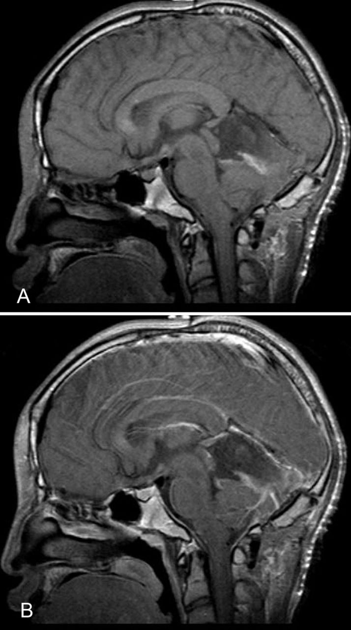
Magnetic resonance imaging (MRI) revealed a solid and cystic enhancing mass centered in the region of the tectum that extended inferiorly to the superior aspect of the fourth ventricle (Figure 1). The lesion measured 3.9 cm × 3.6 cm × 2.4 cm and displaced the posterior midbrain and cerebral aqueduct without complete obstruction. There was slight prominence of the lateral ventricles bilaterally, with enlargement of the temporal horns.

Figure 1.
  • Download figure
  • Open in new tab
  • Download powerpoint
Figure 1.

Magnetic resonance imaging without contrast (A) and with contrast (B) shows a solid and cystic enhancing mass measuring 3.9 cm × 3.6 cm × 2.4 cm centered in the region of the tectum, extending down to the superior aspect of the fourth ventricle with mass effect on the posterior midbrain and displacement of the cerebral aqueduct and with maintained patency.

The patient underwent a suboccipital infratentorial-supracerebellar approach for tumor resection during which an incision from the inion to C2 was outlined for enough exposure. Intraoperative neuronavigation was used to place 2 burr holes just caudal to the transverse sinus bilaterally, followed by a craniotomy. The tumor was best approached by a dissection over the cerebellar hemispheres and an incision on the cerebellar vermis. Grossly, the tumor was yellow-grey and partially collapsed after decompression of the cystic component. The tumor, which extended inferior to the roof of the fourth ventricle, was removed without difficulty using microsurgical technique and ultrasonic aspiration.

Pathology was consistent with PXA (Figure 2). Cytologically, the tumor cells showed marked heterogeneity. In some areas, the tumor cell nuclei were round to slightly oval, with mild to moderate nuclear pleomorphism set in a fibrillary background. In other areas, the nuclei were markedly pleomorphic with occasional multinucleated giant cells. Intranuclear cytoplasmic inclusions were prominent throughout, in addition to a high degree of vascularization. All tumor cells were diffusely and strongly positive for glial fibrillary acidic protein (GFAP). Reticulin stains showed the presence of pericellular fibers, and the Ki-67 proliferation index was 1% to 2%.

Figure 2.
  • Download figure
  • Open in new tab
  • Download powerpoint
Figure 2.

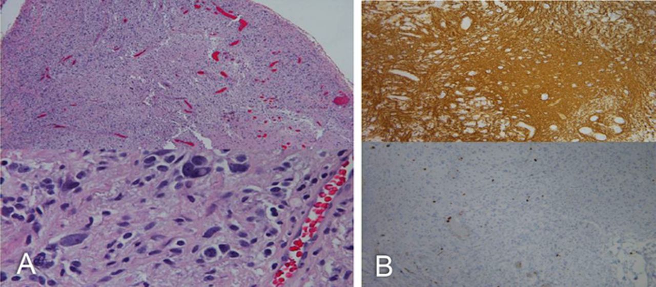
(A) Hematoxylin and eosin stain of the tumor cells shows marked heterogeneity. In some areas, the tumor cells had round to slightly oval nuclei with mild to moderate nuclear pleomorphism set in a fibrillary background. In other areas, the tumor nuclei were markedly pleomorphic with occasional multinucleated giant cells throughout. (B) Intranuclear cytoplasmic inclusions are prominent throughout, in addition to a high degree of vascularization. All tumor cells were diffusely and strongly positive for glial fibrillary acidic protein (top); Ki-67 (bottom) and reticulin stains showed the presence of pericellular fibers. The Ki-67 proliferation index was between 1% and 2%.

Postoperatively, the patient recovered well without any neurologic deficits and was discharged 5 days later. Following discharge, the patient resumed his normal daily activities. Postoperative MRI showed no evidence of residual tumor (Figure 3), and the patient was managed expectantly without the need for adjuvant radiation or chemotherapy. At 4-week, 8-week, 6-month, and 9-month postoperative follow-up, the patient had resolution of symptoms and no evidence of recurrence.

Figure 3.
  • Download figure
  • Open in new tab
  • Download powerpoint
Figure 3.

Magnetic resonance imaging without contrast (A) and with contrast (B) after surgical treatment showed postoperative changes and no evidence of residual tumor.

DISCUSSION

A growing body of literature provides histologic and genetic bases for differentiation of PXA. Such means of differentiation become especially important when PXAs occur in unusual locations, such as the pineal region. In general, tumors of the pineal gland are rare, accounting for 0.4% to 1.0% of all intracranial lesions in adults, 2% to 8% of intracranial neoplasms in children, and <1% of all intracranial tumors overall.4 Primary pineal tumors can be categorized as germ cell tumors, which are most common, or as pineal parenchymal tumors, which are comprised of low-grade pineocytomas, intermediate-grade pineal parenchymal tumors, and highly malignant pineoblastoma.21 Other neoplasms in the pineal region can arise from cell types in the vicinity of the pineal gland such as meningioma, astrocytic tumors, and metastases from distal sites.22 Astrocytic tumors include pilocytic astrocytoma, anaplastic astrocytoma, glioblastoma multiforme, pleomorphic granular cell astrocytoma,23,24 and PXA.

The 4 known cases of PXA of the pineal region are listed in Table 2.4,23,25,26 Each patient presented with signs and symptoms of increased intracranial pressure, including severe headache and papilledema from obstructive hydrocephalus. The patient with the PXA that underwent anaplastic transformation additionally presented with dementia and gait disturbance.25 Our patient's blurred vision and headaches were mild compared to the other cases; because of the heightened vigilance for intracranial tumors in patients with NF1, his diagnosis was made early before he developed obstructive hydrocephalus.

View this table:
  • View inline
  • View popup
Table 2.

Pleomorphic Xanthoastrocytomas of the Pineal Region

Generally, surgical resection of PXAs is curative; however, malignant or anaplastic characteristics are associated with recurrence and poor outcome and may require radiation, chemotherapy, or other adjuvant therapies.11,12,19 Whether an association with NF1 portends a different prognosis for PXAs is uncertain. Of 15 NF1-associated PXAs with a known classification reported in the literature, 4 tumors were anaplastic and 2 were malignant. NF1-associated PXAs, therefore, appear to have worse outcomes than PXAs in the general population, as the malignant or anaplastic transformation in the general population is estimated to be 10% to 20%.19 Accurate diagnosis of PXAs in patients with NF1 is essential and should be considered in atypical presentations or locations.

Diagnosing a pineal PXA in the setting of NF1 is challenging, especially considering the plethora of other tumors more commonly associated with NF1. Histologic characteristics include eosinophilic granules, xanthomatous changes, and nuclear and cellular pleomorphism.27 Because of their histologic similarity to other tumors, such as those of the pineal region, PXAs are differentiated by immunohistochemical, structural, and genetic markers.27 For example, PXAs share a bizarre morphology with the malignant and aggressive giant cell glioblastoma multiforme28,29 that may also occur in the pineal region.14 Our patient's tumor was notably heterogeneous as it showed both marked and moderate pleomorphism in different areas, a high degree of vascularization, and multinucleated giant cells. Although these characteristics are found in glioblastoma multiforme, that diagnosis was ruled out on the basis of the low Ki-67 index and the absence of mitoses or necrosis. Another diagnostic possibility was pleomorphic granular cell tumor. Both tumors are rarely found in the pineal region, they share a similarly indolent course, and their foundational histology shows lipidized pleomorphic granular cells.24,25 However, pleomorphic granular cell tumors show an absence of reticulin fibers and the presence of large numbers of mitochondria,4 which is incongruent with our pathologic findings. To further distinguish between the 2 tumor types, PXAs show diffuse GFAP positivity compared to an occasional weakly positive GFAP positivity in pleomorphic granular cell tumors. Our pathologic specimen was diffusely positive for GFAP and stained positive for pericellular reticulin fibers.

Despite advances in immunohistochemical analysis, distinguishing PXA from pilocytic astrocytoma, which is the most common intracranial tumor associated with NF1, can be challenging. Koelsche et al found the presence of a BRAF V600E mutation in 70% of PXAs, principally within the temporal lobe, and proposed a common molecular mechanism of abnormal Raf kinase activity for both NF1-associated lesions and NF1-associated PXAs.30 A study by Jacob et al also suggested a possible association between BRAF V600E gene mutations and pilocytic astrocytoma.31 In contrast are the 2 reports of BRAF negativity in NF1-associated PXAs.16,17 Because of the historic lack of BRAF testing in NF1-associated PXAs, BRAF significance will remain unclear until larger studies are analyzed. In the meantime, morphologic features will continue to be used to distinguish PXAs from pilocytic astrocytomas. BRAF mutation has also been suggested as a marker of progression-free survival in PXAs,32 and salvage therapy with the BRAF inhibitor vemurafenib for recurrent PXAs has shown some success.33 Although we did not analyze BRAF and other genetic mutations in our case, we advocate for BRAF analysis in all cases of PXA, especially NF1-associated PXAs. Establishing the link between BRAF mutations and PXAs has the potential to provide a more comprehensive understanding of etiology, diagnosis, and treatment.

In a 2019 study of 19 patients with PXA of which 15 underwent anaplastic progression, Phillips et al identified a combination of CDKN2A biallelic deletion and an Raf alteration in all patients in the cohort, suggesting a specific genetic profile, as well as an indicator for anaplastic progression in PXA.34 Furthermore, 47% of anaplastic PXAs were found to have a telomerase reverse transcriptase (TERT) gene alteration. This study and others may further elucidate the role of TERT alterations in PXAs, as that role has not been well defined.35

CONCLUSION

PXAs are rare, WHO grade II brain neoplasms that are rarely associated with NF1. Although PXAs are predominantly temporal lobe–based lesions, they have been found in atypical locations such as the pineal region. To our knowledge, we report only the fifth case of a pineal region PXA and the first case in a patient with NF1. Although the literature is scarce, NF1-associated PXAs may portend a higher degree of malignancy and poorer outcomes compared to PXAs that are not associated with NF1; therefore, diagnostic vigilance via histopathologic and immunohistochemical analyses is imperative. Distinguishing PXA from its similar glial counterparts is also important. Genetic mutations may not only help in diagnosis but also potentially help us understand tumor genesis and provide new targets for treatment. PXA should be included in the differential for NF1-associated intracranial lesions, regardless of location.

This article meets the Accreditation Council for Graduate Medical Education and the American Board of Medical Specialties Maintenance of Certification competencies for Patient Care and Medical Knowledge.

ACKNOWLEDGMENTS

The authors have no financial or proprietary interest in the subject matter of this article.

  • ©2020 by the author(s); Creative Commons Attribution License (CC BY)

©2020 by the author(s); licensee Ochsner Journal, Ochsner Clinic Foundation, New Orleans, LA. This article is an open access article distributed under the terms and conditions of the Creative Commons Attribution (CC BY) license (creativecommons.org/licenses/by/4.0/legalcode) that permits unrestricted use, distribution, and reproduction in any medium, provided the original author(s) and source are credited.

REFERENCES

  1. 1.↵
    1. Kepes JJ,
    2. Rubinstein LJ,
    3. Eng LF
    . Pleomorphic xanthoastrocytoma: a distinctive meningocerebral glioma of young subjects with relatively favorable prognosis. A study of 12 cases. Cancer. 1979 Nov;44(5):1839-1852.
    OpenUrlCrossRefPubMed
  2. 2.↵
    1. Zarate JO,
    2. Sampaolesi R
    . Pleomorphic xanthoastrocytoma of the retina. Am J Surg Pathol. 1999 Jan;23(1):79-81.
    OpenUrlCrossRefPubMed
  3. 3.↵
    1. Herpers MJ,
    2. Freling G,
    3. Beuls EA
    . Pleomorphic xanthoastrocytoma in the spinal cord. Case report. J Neurosurg. 1994 Mar;80(3):564-569. doi: 10.3171/jns.1994.80.3.0564.
    OpenUrlCrossRefPubMed
  4. 4.↵
    1. Srinivas BH,
    2. Uppin MS,
    3. Panigrahi MK,
    4. Vijaya Saradhi M,
    5. Jyotsna Rani Y,
    6. Challa S
    . Pleomorphic xanthoastrocytoma of the pineal region. J Clin Neurosci. 2010;17:1439-1441. doi: 10.1016/j.jocn.2010.02.022.
    OpenUrlCrossRefPubMed
  5. 5.↵
    1. Pasmant E,
    2. Parfait B,
    3. Luscan A,
    4. et al.
    Neurofibromatosis type 1 molecular diagnosis: what can NGS do for you when you have a large gene with loss of function mutations? Eur J Hum Genet. 2015 May;23(5):596-601. doi: 10.1038/ejhg.2014.145.
    OpenUrlCrossRefPubMed
  6. 6.↵
    1. Ozek MM,
    2. Sav A,
    3. Pamir MN,
    4. Ozer AF,
    5. Ozek E,
    6. Erzen C
    . Pleomorphic xanthoastrocytoma associated with von Recklinghausen neurofibromatosis. Childs Nerv Syst. 1993 Feb;9(1):39-42.
    OpenUrlCrossRefPubMed
  7. 7.
    1. Kubo O,
    2. Sasahara A,
    3. Tajika Y,
    4. Kawamiira H,
    5. Kawabatake H,
    6. Takakura K
    . Pleomorphic xanthoastrocytoma with neurofibromatosis type 1: case report. Noshuyo Byori. 1996 Apr;13(1):79-83.
    OpenUrlPubMed
  8. 8.
    1. Koeller KK,
    2. Henry JM
    . From the archives of the AFIP: superficial gliomas: radiologic-pathologic correlation. Armed Forces Institute of Pathology. Radiographics. 2001 Nov-Dec;21(6):1533-1556. doi: 10.1148/radiographics.21.6.g01nv051533.
    OpenUrlCrossRefPubMed
  9. 9.
    1. Naidich MJ,
    2. Walker MT,
    3. Gottardi-Littelt NR,
    4. Han G,
    5. Chandler JP
    . Cerebellar pleomorphic xanthoastrocytoma in a patient with neurofibromatosis type 1. Neuroradiology. 2004 Oct;46(10):825-829. doi: 10.1007/s00234-004-1216-0.
    OpenUrlCrossRefPubMed
  10. 10.
    1. Saikali S,
    2. Le Strat A,
    3. Heckly A,
    4. Stock N,
    5. Scarabin JM,
    6. Hamlat A
    . Multicentric pleomorphic xanthoastrocytoma in a patient with neurofibromatosis type 1. Case report and review of the literature. J Neurosurg. 2005 Feb;102(2):376-381. doi: 10.3171/jns.2005.102.2.0376.
    OpenUrlCrossRefPubMed
  11. 11.↵
    1. Hariharan S,
    2. Donahue JE,
    3. Garre C,
    4. Origone P,
    5. Grewal RP
    . Clinicopathologic and genetic analysis of siblings with NF1 and adult-onset gliomas. J Neurol Sci. 2006 Aug 15;247(1):105-108. doi: 10.1016/j.jns.2006.03.020.
    OpenUrlCrossRefPubMed
  12. 12.↵
    1. Horiguchi S,
    2. Mitsuya K,
    3. Watanabe R,
    4. Yagishita S,
    5. Nakasu Y
    . Pleomorphic xanthoastrocytoma and moyamoya disease in a patient with neurofibromatosis type 1 – case report. Neurol Med Chir (Tokyo). 2011;51(4):310-314.
    OpenUrlPubMed
  13. 13.
    1. Adeleye AO,
    2. Okolo CA,
    3. Akang EE,
    4. Adesina AM
    . Cerebral pleomorphic xanthoastrocytoma associated with NF1: an updated review with a rare atypical case from Africa. Neurosurg Rev. 2012 Jul;35(3):313-319; discussion 319. doi: 10.1007/s10143-011-0362-1.
    OpenUrlCrossRefPubMed
  14. 14.↵
    1. Neal MT,
    2. Ellis TL,
    3. Stanton CA
    . Pleomorphic xanthoastrocytoma in two siblings with neurofibromatosis type 1 (NF1). Clin Neuropathol. 2012 Jan-Feb;31(1):54-56. doi: 10.5414/NP300424.
    OpenUrlCrossRefPubMed
  15. 15.
    1. Prayson RA
    . Pleomorphic xanthoastrocytoma arising in neurofibromatosis type 1. Clin Neuropathol. 2012 May-Jun;31(3):152-154.
    OpenUrlCrossRefPubMed
  16. 16.↵
    1. Vizcaino MA,
    2. Caccamo DV,
    3. Fox E,
    4. Rodriguez FJ
    . Pleomorphic xanthoastrocytoma: report of two cases with unconventional clinical presentations. Clin Neuropathol. 2014 Nov-Dec;33(6):380-387. doi: 10.5414/NP300766.
    OpenUrlCrossRef
  17. 17.↵
    1. Takei H,
    2. Rouah E,
    3. Bhattacharjee MB
    . Cerebellar pleomorphic xanthoastrocytoma in a patient with neurofibromatosis type 1: a case report and literature review. Int J Clin Exp Pathol. 2015 Jun 1;8(6):7570-7574.
    OpenUrlPubMed
  18. 18.
    1. Thara K,
    2. Sharma R,
    3. Thiagarajan G,
    4. Ramdas A,
    5. Varghese RG
    . Anaplastic pleomorphic xanthoastrocytoma in a case of neurofibromatosis type 1: a case report. J Clin Diagn Res. 2017 Apr;11(4):ED23-ED24. doi: 10.7860/JCDR/2017/26685.9713.
    OpenUrlCrossRef
  19. 19.↵
    1. Singla N,
    2. Kapoor A,
    3. Radotra BD,
    4. Chatterjee D
    . Malignant conversion to glioblastoma in neurofibromatosis type I-associated pleomorphic xanthoastrocytoma: unknown predictors of favorable outcome. Asian J Neurosurg. 2018 Jul-Sep;13(3):826-829.
    OpenUrl
  20. 20.↵
    1. Louis DN,
    2. Perry A,
    3. Reifenberger G,
    4. et al.
    The 2016 World Health Organization classification of tumors of the central nervous system: a summary. Acta Neuropathol. 2016 Jun; 131(6): 803-820. doi: 10.1007/s00401-016-1545-1.
    OpenUrlCrossRefPubMed
  21. 21.↵
    1. Louis DN,
    2. Ohgaki H,
    3. Wiestler OD,
    4. et al.
    The 2007 WHO classification of tumours of the central nervous system. Acta Neuropathol. 2007 Aug;114(2):97-109. doi: 10.1007/s00401-007-0243-4. Erratum in: Acta Neuropathol. 2007. November;114(5): 547.
    OpenUrlCrossRefPubMed
  22. 22.↵
    1. Inoue T,
    2. Kumabe T,
    3. Kanamori M,
    4. Sonoda Y,
    5. Watanabe M,
    6. Tominaga T
    . Papillary tumor of the pineal region: a case report. Brain Tumor Pathol. 2008;25(2):85-90. doi: 10.1007/s10014-008-0231-y.
    OpenUrlCrossRefPubMed
  23. 23.↵
    1. Ohta T,
    2. Yachi K,
    3. Ogino A,
    4. et al.
    Pleomorphic granular cell astrocytoma in the pineal gland: case report. Neuropathology. 2010 Dec;30(6):615-620. doi: 10.1111/j.1440-1789.2009.01095.x.
    OpenUrlCrossRefPubMed
  24. 24.↵
    1. Snipes GJ,
    2. Horoupian DS,
    3. Shuer LM,
    4. Silverberg GD
    . Pleomorphic granular cell astrocytoma of the pineal gland. Cancer. 1992 Oct 15;70(8):2159-2165.
    OpenUrlPubMed
  25. 25.↵
    1. Katayama K,
    2. Asano K,
    3. Shimamura N,
    4. et al.
    Case of pleomorphic xanthoastrocytoma with anaplastic features in the pineal gland. Brain Tumor Pathol. 2013 Oct;30(4):242-246. doi: 10.1007/s10014-013-0137-1.
    OpenUrlCrossRefPubMed
  26. 26.↵
    1. Thakar S,
    2. Sai Kiran NA,
    3. Ghosal N,
    4. Hegde AS
    . Pleomorphic xanthoastrocytoma: a new differential diagnosis for a pediatric pineal neoplasm. Brain Tumor Pathol. 2012 Jul;29(3):168-171. doi: 10.1007/s10014-011-0076-7.
    OpenUrlCrossRefPubMed
  27. 27.↵
    1. Dias-Santagata D,
    2. Lam Q,
    3. Vernovsky K,
    4. et al.
    BRAF V600E mutations are common in pleomorphic xanthoastrocytoma: diagnostic and therapeutic implications. PLoS One. 2011 Mar 29;6(3):e17948. doi: 10.1371/journal.pone.0017948.
    OpenUrlCrossRefPubMed
  28. 28.↵
    1. Scheithauer BW,
    2. Aker AT,
    3. Ketterling RP,
    4. Carlson AW,
    5. Knudson RA,
    6. Tyler M
    . Anaplastic astroblastoma-sarcoma in neurofibromatosis type 1. Clin Neuropathol. 2010 Sep-Oct;29(5):289-296.
    OpenUrlPubMed
  29. 29.↵
    1. Kepes JJ
    . Glioblastoma multiforme masquerading as a pleomorphic xanthoastrocytoma. Childs Nerv Syst. 1989 Jun;5(3):127.
    OpenUrlPubMed
  30. 30.↵
    1. Koelsche C,
    2. Sahm F,
    3. Wöhrer A,
    4. et al.
    BRAF-mutated pleomorphic xanthoastrocytoma is associated with temporal location, reticulin fiber deposition and CD34 expression. Brain Pathol. 2014 Apr; 24(3):221-229. doi: 10.1111/bpa.12111.
    OpenUrlCrossRefPubMed
  31. 31.↵
    1. Jacob K,
    2. Quang-Khuong DA,
    3. Jones DT,
    4. et al.
    Genetic aberrations leading to MAPK pathway activation mediate oncogene-induced senescence in sporadic pilocytic astrocytomas. Clin Cancer Res. 2011 Jul 15;17(14):4650-4660. doi: 10.1158/1078-0432.CCR-11-0127.
    OpenUrlAbstract/FREE Full Text
  32. 32.↵
    1. Tabouret E,
    2. Bequet C,
    3. Denicolai E,
    4. et al.
    BRAF mutation and anaplasia may be predictive factors of progression-free survival in adult pleomorphic xanthoastrocytoma. Eur J Surg Oncol. 2015 Dec;41(12):1685-1690. doi: 10.1016/j.ejso.2015.09.012.
    OpenUrlCrossRefPubMed
  33. 33.↵
    1. Chamberlain MC
    . Salvage therapy with BRAF inhibitors for recurrent pleomorphic xanthoastrocytoma: a retrospective case series. J Neurooncol. 2013 Sep;114(2):237-240. doi: 10.1007/s11060-013-1176-5.
    OpenUrlCrossRefPubMed
  34. 34.↵
    1. Phillips JJ,
    2. Gong H,
    3. Chen K,
    4. et al.
    The genetic landscape of anaplastic pleomorphic xanthoastrocytoma. Brain Pathol. 2019 Jan;29(1):85-96. doi: 10.1111/bpa.12639.
    OpenUrlCrossRef
  35. 35.↵
    1. Ma C,
    2. Feng R,
    3. Chen H,
    4. et al.
    BRAF V600E, TERT, and IDH2 mutations in pleomorphic xanthoastrocytoma: observations from a large case-series study. World Neurosurg. 2018 Dec;120:e1225-e1233. doi: 10.1016/j.wneu.2018.09.050.
    OpenUrlCrossRef
PreviousNext
Back to top

In this issue

Ochsner Journal: 20 (2)
Ochsner Journal
Vol. 20, Issue 2
Jun 2020
  • Table of Contents
  • Table of Contents (PDF)
  • About the Cover
  • Index by author
  • Advertising (PDF)
  • Back Matter (PDF)
  • Ed Board (PDF)
  • Front Matter (PDF)
Print
Download PDF
Email Article

Thank you for your interest in spreading the word on Ochsner Journal.

NOTE: We only request your email address so that the person you are recommending the page to knows that you wanted them to see it, and that it is not junk mail. We do not capture any email address.

Enter multiple addresses on separate lines or separate them with commas.
Pleomorphic Xanthoastrocytoma of the Pineal Region in a Pediatric Patient With Neurofibromatosis Type 1
(Your Name) has sent you a message from Ochsner Journal
(Your Name) thought you would like to see the Ochsner Journal web site.
CAPTCHA
This question is for testing whether or not you are a human visitor and to prevent automated spam submissions.
Citation Tools
Pleomorphic Xanthoastrocytoma of the Pineal Region in a Pediatric Patient With Neurofibromatosis Type 1
Joshua A. Hanna, Mansour Mathkour, Edna E. Gouveia, JonMark Lane, Lauren Boehm, Joseph R. Keen, Erin E. Biro, Olawale A. Sulaiman, Cuong J. Bui
Ochsner Journal Jun 2020, 20 (2) 226-231; DOI: 10.31486/toj.18.0156

Citation Manager Formats

  • BibTeX
  • Bookends
  • EasyBib
  • EndNote (tagged)
  • EndNote 8 (xml)
  • Medlars
  • Mendeley
  • Papers
  • RefWorks Tagged
  • Ref Manager
  • RIS
  • Zotero
Share
Pleomorphic Xanthoastrocytoma of the Pineal Region in a Pediatric Patient With Neurofibromatosis Type 1
Joshua A. Hanna, Mansour Mathkour, Edna E. Gouveia, JonMark Lane, Lauren Boehm, Joseph R. Keen, Erin E. Biro, Olawale A. Sulaiman, Cuong J. Bui
Ochsner Journal Jun 2020, 20 (2) 226-231; DOI: 10.31486/toj.18.0156
Reddit logo Twitter logo Facebook logo Mendeley logo
  • Tweet Widget
  • Facebook Like
  • Google Plus One

Jump to section

  • Article
    • Abstract
    • INTRODUCTION
    • CASE REPORT
    • DISCUSSION
    • CONCLUSION
    • ACKNOWLEDGMENTS
    • REFERENCES
  • Figures & Data
  • Info & Metrics
  • References
  • PDF

Cited By...

  • Systematic discovery of gene fusions in pediatric cancer by integrating RNA-seq and WGS
  • Google Scholar

More in this TOC Section

  • Pediatric Undifferentiated Pleomorphic Sarcoma of the Cecum
  • Differential to Terminal Ileitis: Terminal Ileum Neuroendocrine Tumor Identified on Screening Colonoscopy
  • Pelvic Synovial Sarcoma Clinically Masquerading as an Ovarian Malignancy
Show more CASE REPORTS AND CLINICAL OBSERVATIONS

Similar Articles

Keywords

  • Neurofibromatosis type 1
  • pediatrics
  • pineal region
  • pleomorphic astrocytoma

Current Post at the Blog

No Author Credit for ChatGPT

Our Content

  • Home
  • Current Issue
  • Ahead of Print
  • Archive
  • Featured Contributors
  • Ochsner Journal Blog
  • Archive at PubMed Central

Information & Forms

  • Instructions for Authors
  • Instructions for Reviewers
  • Submission Checklist
  • FAQ
  • License for Publishing-Author Attestation
  • Patient Consent Form
  • Submit a Manuscript

Services & Contacts

  • Permissions
  • Sign up for our electronic table of contents
  • Feedback Form
  • Contact Us

About Us

  • Editorial Board
  • About the Ochsner Journal
  • Ochsner Health
  • University of Queensland-Ochsner Clinical School
  • Alliance of Independent Academic Medical Centers

© 2023 Ochsner Clinic Foundation

Powered by HighWire