
Main menu

  • Home
  • Content
    • Current
    • Ahead of print
    • Archive
  • Info for
    • Authors
    • Reviewers
  • About Us
    • About the Ochsner Journal
    • Editorial Board
  • More
    • Alerts
    • Feedback
  • Other Publications
    • Ochsner Journal Blog

User menu

  • My alerts
  • Log in

Search

  • Advanced search
Ochsner Journal
  • Other Publications
    • Ochsner Journal Blog
  • My alerts
  • Log in
Ochsner Journal

Advanced Search

  • Home
  • Content
    • Current
    • Ahead of print
    • Archive
  • Info for
    • Authors
    • Reviewers
  • About Us
    • About the Ochsner Journal
    • Editorial Board
  • More
    • Alerts
    • Feedback
Case ReportCASE REPORTS AND CLINICAL OBSERVATIONS
Open Access

Collision Tumor of the Kidney: Renal Cell Carcinoma Hidden in a Giant Angiomyolipoma in a Patient With Tuberous Sclerosis Complex

José Iván Robles-Torres, José Gustavo Arrambide-Herrera, Max Molina-Ayala, Sergio Alan Dávila-Martínez, Pedro Antonio Madero-Morales and Lauro Salvador Gómez-Guerra
Ochsner Journal September 2021, 21 (3) 287-290; DOI: https://doi.org/10.31486/toj.20.0075
José Iván Robles-Torres
1Department of Urology, Hospital Universitario Dr. José Eleuterio González, Universidad Autónoma de Nuevo León, Monterrey, Nuevo León, México
MD
  • Find this author on Google Scholar
  • Find this author on PubMed
  • Search for this author on this site
José Gustavo Arrambide-Herrera
1Department of Urology, Hospital Universitario Dr. José Eleuterio González, Universidad Autónoma de Nuevo León, Monterrey, Nuevo León, México
MD
  • Find this author on Google Scholar
  • Find this author on PubMed
  • Search for this author on this site
Max Molina-Ayala
2Department of Pathological Anatomy, Hospital Universitario Dr. José Eleuterio González, Universidad Autónoma de Nuevo León, Monterrey, Nuevo León, México
MD
  • Find this author on Google Scholar
  • Find this author on PubMed
  • Search for this author on this site
Sergio Alan Dávila-Martínez
1Department of Urology, Hospital Universitario Dr. José Eleuterio González, Universidad Autónoma de Nuevo León, Monterrey, Nuevo León, México
MD
  • Find this author on Google Scholar
  • Find this author on PubMed
  • Search for this author on this site
Pedro Antonio Madero-Morales
1Department of Urology, Hospital Universitario Dr. José Eleuterio González, Universidad Autónoma de Nuevo León, Monterrey, Nuevo León, México
MD
  • Find this author on Google Scholar
  • Find this author on PubMed
  • Search for this author on this site
Lauro Salvador Gómez-Guerra
1Department of Urology, Hospital Universitario Dr. José Eleuterio González, Universidad Autónoma de Nuevo León, Monterrey, Nuevo León, México
PhD
  • Find this author on Google Scholar
  • Find this author on PubMed
  • Search for this author on this site
  • For correspondence: laurogomez@hotmail.com
  • Article
  • Figures & Data
  • References
  • Info & Metrics
  • PDF
Loading

Abstract

Background: A renal angiomyolipoma is a mixed mesenchymal benign tumor composed of smooth muscle, adipose tissue, and blood vessels. Malignant transformation of angiomyolipomas is anecdotal. To our knowledge, only 6 cases have been reported, and 4 of the patients had tuberous sclerosis complex diagnosed.

Case Report: We present the case of a 29-year-old male with tuberous sclerosis complex who arrived at the emergency room with gross hematuria and a painful right-sided abdominal mass. Imaging studies revealed active bleeding from a giant angiomyolipoma. An emergency nephrectomy was performed. Histopathology evaluation revealed an angiomyolipoma with a focal lesion and clear cell renal carcinoma within the tumor.

Conclusion: Limited evidence is available to dictate management of collision tumors of the kidney in the scenario of tuberous sclerosis complex, so a multidisciplinary approach that includes urology, oncology, genetics, and nephrology intervention needs to be considered. No standardized follow-up modality has been established for angiomyolipomas, so patients should be placed under active surveillance, similar to that carried out in cases of renal cell carcinoma.

Keywords:
  • Angiomyolipoma
  • carcinoma–renal cell
  • tuberous sclerosis

INTRODUCTION

Tuberous sclerosis complex (TSC)—a rare, multisystemic genetic disorder that causes benign tumors to grow in different organs, such as the brain, eyes, kidneys, heart, lungs, and skin—is the second most common neurocutaneous syndrome with a prevalence rate of 1/6,000 births.1 TSC is caused by mutations on 2 genes: TSC1 and TSC2.2 The most affected organ is the brain, resulting in clinical manifestations such as seizures, mental retardation, and skin abnormalities. The kidney is involved in approximately 80% to 85% of cases.3 Patients with TSC have an increased incidence of tumors, such as multiple renal angiomyolipomas, renal cell carcinoma, and oncocytoma.4 An angiomyolipoma is a mixed mesenchymal benign tumor composed of smooth muscle, adipose tissue, and blood vessels.5 Malignant transformation of these tumors is anecdotal. The coexistence of renal cell carcinoma and renal angiomyolipoma within the same tumor mass, also called collision tumor, is unusual, and, to our knowledge, only 6 cases have been reported.3,4,6-9 We present a case of a young male with TSC and a giant angiomyolipoma that contained a focal lesion with renal cell carcinoma.

CASE REPORT

A 29-year-old male with a medical history of TSC diagnosed at age 16 years presented with sudden onset of gross hematuria and a painful, firm, right-sided abdominal mass. Vital signs upon arrival were temperature of 36.4 °C, blood pressure of 90/60 mmHg, respiration of 22/min, and heart rate of 110/min. Physical examination revealed generalized pallor, multiple cutaneous nodules on the face, hypopigmented skin lesions, and generalized angiofibromas. Abdominal examination showed a painful solid mass located on the right flank. Laboratory findings on arrival were hemoglobin 4.0 g/dL, leukocyte count 14.0 × 10⁹/L, C-reactive protein 4.5 mg/L, creatinine 2.3 mg/dL, and estimated glomerular filtration rate 35.9 mL/min/1.73 m2. Initial resuscitation with 2 L of 0.9% saline solution and 2 units of red blood cells was performed. Transurethral catheter was placed and showed gross hematuria.

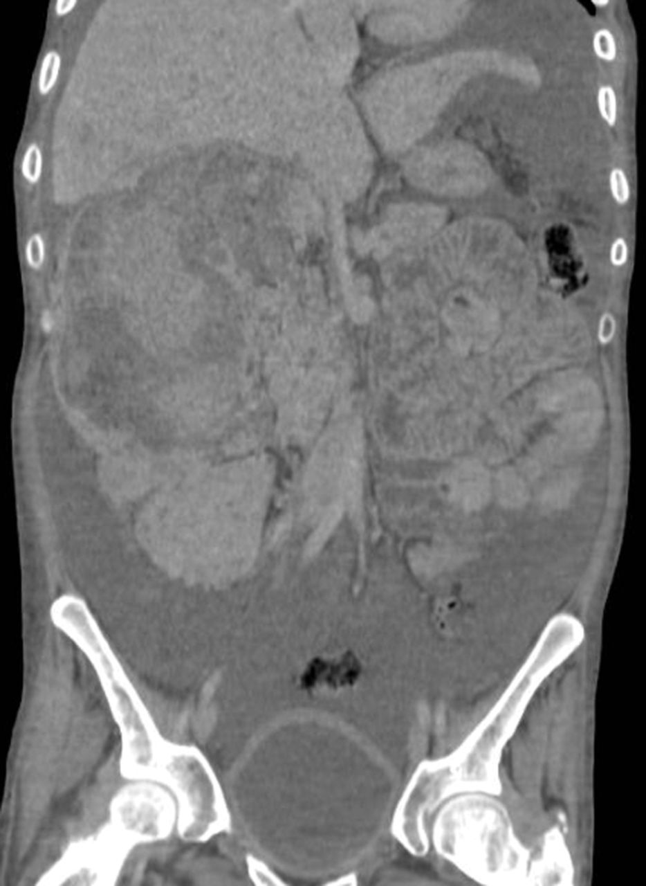
Contrast-enhanced computed tomography (CT) scan showed bilateral renal angiomyolipomas, with a predominant right renal mass measuring 12 × 12 × 18 cm3 (Figure 1) and showing signs of active arterial bleeding. A simple nephrectomy was performed. The kidney was enlarged and irregular, with heterogeneous tanning and admixed fat tissue within the tumor (Figure 2). Light microscopy showed multiple angiomyolipomas, and one section showed a well-circumscribed lesion within the giant angiomyolipoma that was composed of clear cells with finely granular eosinophilic cytoplasm and marked nuclear enlargement with prominent nucleoli (Figure 3).

Figure 1.
  • Download figure
  • Open in new tab
  • Download powerpoint
Figure 1.

Computed tomography scan in coronal view shows a large right perinephric hematoma, left kidney with multiple angiomyolipomas, and abundant free fluid in the peritoneal cavity.

Figure 2.
  • Download figure
  • Open in new tab
  • Download powerpoint
Figure 2.

(A) Macroscopic view of the kidney. (B) Cross-section of the kidney shows visible tumor with a tan, whorled appearance and admixed fat tissue (asterisk). The ureter was identified (arrow).

Figure 3.
  • Download figure
  • Open in new tab
  • Download powerpoint
Figure 3.

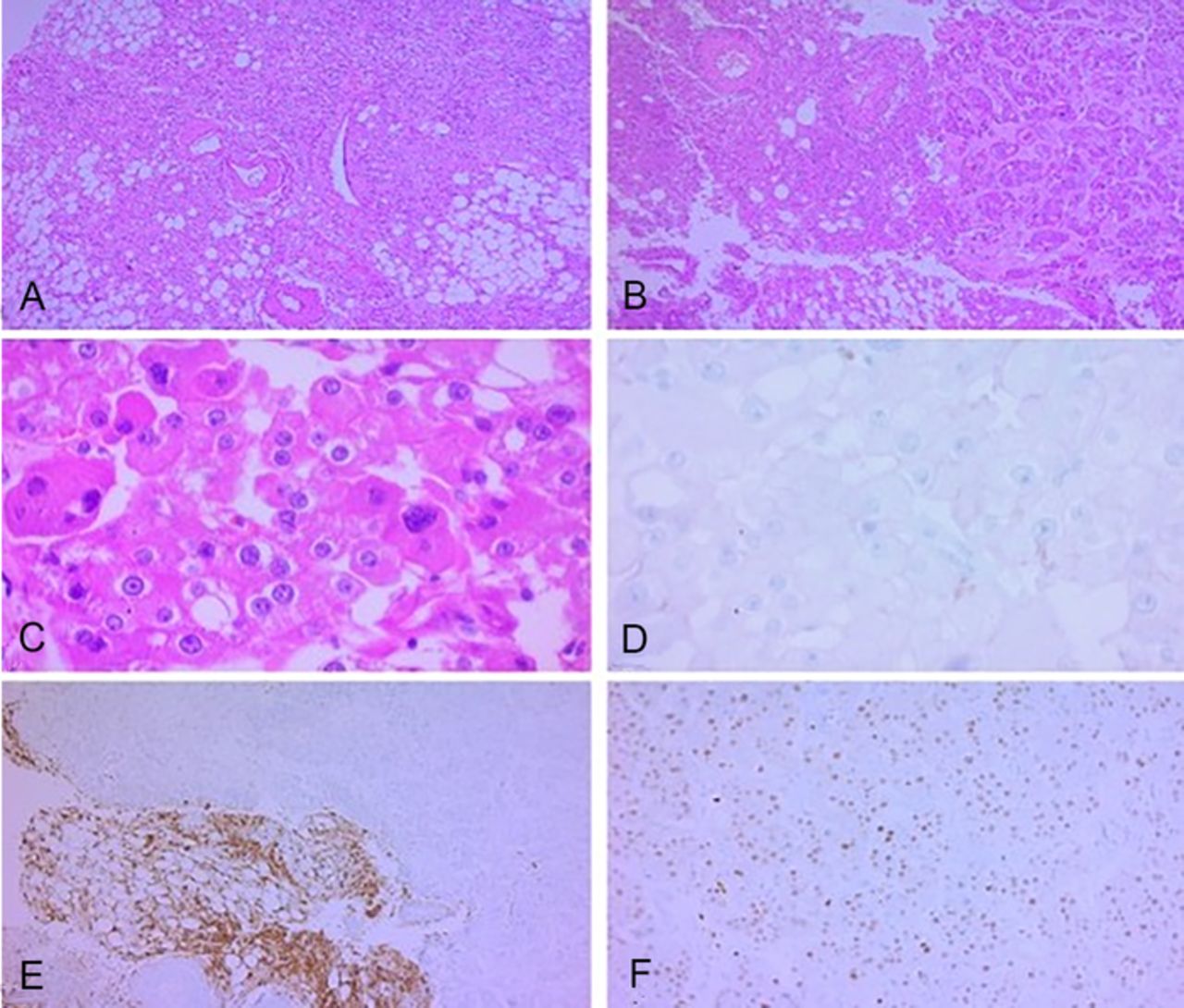
Hematoxylin and eosin stained sections of the patient's kidney. (A) The arrangement of adipocytes, spindle cells, and enlarged thick-walled vessels with irregular walls is mostly haphazard (×100). (B) A clear transition to a different tumor is observed in nests within a collagenous stroma (×100). (C) In detail (×400), pleomorphic nuclei and prominent nucleoli are observed, with a finely granular eosinophilic cytoplasm and a fine capillary network, compatible with renal cell carcinoma. The entire area with these characteristics was surrounded by angiomyolipoma. Immunohistochemistry was performed to further identify this foci, with CD117 negativity (D), HMB45 positivity only in the angiomyolipoma component (E), and PAX8 nuclear positivity in the renal cell carcinoma (F).

Immunohistochemistry results were HMB45 positive and PAX8 negative, confirming the diagnosis of angiomyolipoma. The focal lesion had PAX8 positive and HMB45 negative cells, compatible with a renal cell carcinoma clear cell variant, and was completely surrounded by angiomyolipoma. The tumor was classified as World Health Organization/International Society of Urological Pathology (WHO/ISUP) grade 4.

During the postoperative period, the patient's clinical response was favorable, with maintenance of hemodynamic stability. However, his renal function remained affected, and he required temporary renal replacement therapy. After surgery, the patient maintained a baseline creatinine of 1.3 mg/dL and a glomerular filtration rate of 69.4 mL/min/1.73 m2. After a 14-day hospital stay, the patient was discharged with close follow-up.

Surveillance of the contralateral kidney with a multidisciplinary approach involving various specialties—including urology, oncology, radiology, genetics, and nephrology—was chosen because no evidence supports active treatment in this specific setting. Based on the recommendations for active surveillance for renal cell carcinoma, a CT scan was scheduled for 3 months after surgery, and then every year if no suspicious changes were detected. Renal biopsy was suggested if a suspicious lesion were detected during follow-up.10 After 1 year of follow-up with periodic clinical and radiologic evaluations, no evidence of recurrence has been reported in the remaining kidney.

DISCUSSION

Coexistence of renal cell carcinoma and angiomyolipoma within the same tumor is anecdotal, with only 6 cases reported,3,4,6-9 and 4 of the patients had TSC.3,4,6,8 The mean age of the patients with collision tumors was 25.5 ± 1.2 years, similar to our case.3,4,6-9 Approximately 50% to 80% of patients with TSC will have angiomyolipomas.4 Renal angiomyolipomas are mixed mesenchymal tumors belonging to the family of perivascular epithelioid cell tumors; they are the most common kidney manifestation in TSC.5,11 In this scenario, angiomyolipomas are bilateral, multiple, and usually asymptomatic. In some cases, angiomyolipomas can have an accelerated growth rate, causing pain, kidney failure, or sudden bleeding.11

Even in cases with cytologic atypia or lymph node involvement, angiomyolipomas are considered benign. In cases of transformation to renal cell carcinoma variants, the initial morphology mandates are supported with immunohistochemistry. Angiomyolipomas are positive for melanocytic tumor markers, such as HMB45 and Melan-A, and can also be expressed in mature fat tissue and blood vessels.12 Granular eosinophilic cytoplasm warrants the differential diagnosis of oncocytoma, clear cell, and chromophobe variants of renal cell carcinoma. Some of the most important and useful markers for differential diagnosis of renal cell carcinoma are cytokeratins, vimentin, PAX2, and PAX8. Renal cell carcinoma is negative for melanocytic tumor markers.12

No standardized follow-up has been established for angiomyolipomas. Indications for further imaging studies are angiomyolipomas >4 cm, recurrent hematuria, and pain related to tumor growth. Once renal cell carcinoma is confirmed after nephrectomy, a CT scan must be performed 3 months after surgery and then annually for 3 years or longer as clinically indicated. A more rigorous imaging schedule can be considered if positive margins or adverse pathologic features are reported, such as sarcomatoid morphology or high grade (3 or 4) according to the WHO/ISUP staging system.10

CONCLUSION

Malignant transformation of angiomyolipomas is extremely rare and limited to anecdotal reports. Most of the patients reported with these findings have TSC. Because of limited evidence, an etiologic cause of this condition cannot be established. Active surveillance is recommended in patients with TSC and angiomyolipomas, similar to the surveillance recommended for renal cell carcinoma, with the aim of detecting early changes suggestive of malignant transformation or complications related to tumor growth.

This article meets the Accreditation Council for Graduate Medical Education and the American Board of Medical Specialties Maintenance of Certification competencies for Patient Care and Medical Knowledge.

ACKNOWLEDGMENTS

Because of the patient's cognitive condition, parents gave their written informed consent to publish this case, including images of the surgical specimen and histopathologic study. The authors have no financial or proprietary interest in the subject matter of this article.

  • ©2021 by the author(s); Creative Commons Attribution License (CC BY)

©2021 by the author(s); licensee Ochsner Journal, Ochsner Clinic Foundation, New Orleans, LA. This article is an open access article distributed under the terms and conditions of the Creative Commons Attribution (CC BY) license (creativecommons.org/licenses/by/4.0/legalcode) that permits unrestricted use, distribution, and reproduction in any medium, provided the original author(s) and source are credited.

REFERENCES

  1. 1.↵
    1. Curatolo P,
    2. Verdecchia M,
    3. Bombardieri R
    . Tuberous sclerosis complex: a review of neurological aspects. Eur J Paediatr Neurol. 2002;6(1):15-23. doi: 10.1053/ejpn.2001.0538
    OpenUrlCrossRefPubMed
  2. 2.↵
    1. Northrup H,
    2. Krueger DA
    ; International Tuberous Sclerosis Complex Consensus Group. Tuberous sclerosis complex diagnostic criteria update: recommendations of the 2012 International Tuberous Sclerosis Complex Consensus Conference. Pediatr Neurol. 2013;49(4):243-254. doi: 10.1016/j.pediatrneurol.2013.08.001
    OpenUrlCrossRefPubMed
  3. 3.↵
    1. Wang CN,
    2. Wang SY,
    3. Lin MJ,
    4. Wang HW
    . Occult renal cell carcinoma of eosinophilic morphology detected within renal angiomyolipoma mass in a patient with tuberous sclerosis complex. Chin Med J (Engl). 2017;130(12):1505-1506. doi: 10.4103/0366-6999.207476
    OpenUrlCrossRef
  4. 4.↵
    1. Lynne CM,
    2. Carrion HM,
    3. Bakshandeh K,
    4. Nadji M,
    5. Russel E,
    6. Politano VA
    . Renal angiomyolipoma; polycystic kidney, and renal cell carcinoma in a patient with tuberous sclerosis. Urology. 1979;14(2):174-176. doi: 10.1016/0090-4295(79)90153-5
    OpenUrlCrossRefPubMed
  5. 5.↵
    1. Stone CH,
    2. Lee MW,
    3. Amin MB,
    4. et al.
    Renal angiomyolipoma: further immunophenotypic characterization of an expanding morphologic spectrum. Arch Pathol Lab Med. 2001;125(6):751-758.
    OpenUrlPubMed
  6. 6.↵
    1. Taylor RS,
    2. Joseph DB,
    3. Kohaut EC,
    4. Wilson ER,
    5. Bueschen AJ
    . Renal angiomyolipoma associated with lymph node involvement and renal cell carcinoma in patients with tuberous sclerosis. J Urol. 1989;141(4):930-932. doi: 10.1016/S0022-5347(17)41057-3
    OpenUrlCrossRefPubMed
  7. 7.
    1. Huang JK,
    2. Ho DM,
    3. Wang JH,
    4. Chou YH,
    5. Chen MT,
    6. Chang SS
    . Coincidental angiomyolipoma and renal cell carcinoma—report of 1 case and review of literature. J Urol. 1988;140(6):1516-1518. doi: 10.1016/S0022-5347(17)42091-X
    OpenUrlCrossRefPubMed
  8. 8.↵
    1. Schujman E,
    2. Meiraz D,
    3. Liban E,
    4. Servadio C
    . Mixed renomedullary tumor: renal cell carcinoma associated with angiomyolipoma. Urology. 1981;17(4):375-376. doi: 10.1016/0090-4295(81)90274-0
    OpenUrlCrossRefPubMed
  9. 9.↵
    1. Inomoto C,
    2. Umemura S,
    3. Sasaki Y,
    4. Yasuda M,
    5. Terachi T,
    6. Osamura RY
    . Renal cell carcinoma arising in a long pre-existing angiomyolipoma. Pathol Int. 2007;57(3):162-166. doi: 10.1111/j.1440-1827.2006.02075.x
    OpenUrlCrossRefPubMed
  10. 10.↵
    1. Motzer RJ,
    2. Jonasch E,
    3. Michaelson MD,
    4. et al.
    NCCN guidelines insights: kidney cancer, version 2.2020: featured updates to the NCCN guidelines. J Natl Compr Canc Netw. 2019;17(11):1278-1285. doi 10.6004/jnccn.2019.0054
    OpenUrlCrossRefPubMed
  11. 11.↵
    1. Rakowski SK,
    2. Winterkorn EB,
    3. Paul E,
    4. Steele DJR,
    5. Halpern EF,
    6. Thiele EA
    . Renal manifestations of tuberous sclerosis complex: incidence, prognosis, and predictive factors. Kidney Int. 2006;70(10):1777-1782. doi: 10.1038/sj.ki.5001853
    OpenUrlCrossRefPubMed
  12. 12.↵
    1. Aydin H,
    2. Magi-Galluzzi C,
    3. Lane BR,
    4. et al.
    Renal angiomyolipoma: clinicopathologic study of 194 cases with emphasis on the epithelioid histology and tuberous sclerosis association. Am J Surg Pathol. 2009;33(2):289-297. doi: 10.1097/PAS.0b013e31817ed7a6
    OpenUrlCrossRefPubMed
PreviousNext
Back to top

In this issue

Ochsner Journal: 21 (3)
Ochsner Journal
Vol. 21, Issue 3
Sep 2021
  • Table of Contents
  • Table of Contents (PDF)
  • About the Cover
  • Index by author
  • Advertising (PDF)
  • Back Matter (PDF)
  • Ed Board (PDF)
  • Front Matter (PDF)
Print
Download PDF
Email Article

Thank you for your interest in spreading the word on Ochsner Journal.

NOTE: We only request your email address so that the person you are recommending the page to knows that you wanted them to see it, and that it is not junk mail. We do not capture any email address.

Enter multiple addresses on separate lines or separate them with commas.
Collision Tumor of the Kidney: Renal Cell Carcinoma Hidden in a Giant Angiomyolipoma in a Patient With Tuberous Sclerosis Complex
(Your Name) has sent you a message from Ochsner Journal
(Your Name) thought you would like to see the Ochsner Journal web site.
CAPTCHA
This question is for testing whether or not you are a human visitor and to prevent automated spam submissions.
Citation Tools
Collision Tumor of the Kidney: Renal Cell Carcinoma Hidden in a Giant Angiomyolipoma in a Patient With Tuberous Sclerosis Complex
José Iván Robles-Torres, José Gustavo Arrambide-Herrera, Max Molina-Ayala, Sergio Alan Dávila-Martínez, Pedro Antonio Madero-Morales, Lauro Salvador Gómez-Guerra
Ochsner Journal Sep 2021, 21 (3) 287-290; DOI: 10.31486/toj.20.0075

Citation Manager Formats

  • BibTeX
  • Bookends
  • EasyBib
  • EndNote (tagged)
  • EndNote 8 (xml)
  • Medlars
  • Mendeley
  • Papers
  • RefWorks Tagged
  • Ref Manager
  • RIS
  • Zotero
Share
Collision Tumor of the Kidney: Renal Cell Carcinoma Hidden in a Giant Angiomyolipoma in a Patient With Tuberous Sclerosis Complex
José Iván Robles-Torres, José Gustavo Arrambide-Herrera, Max Molina-Ayala, Sergio Alan Dávila-Martínez, Pedro Antonio Madero-Morales, Lauro Salvador Gómez-Guerra
Ochsner Journal Sep 2021, 21 (3) 287-290; DOI: 10.31486/toj.20.0075
del.icio.us logo Twitter logo Facebook logo Mendeley logo
  • Tweet Widget
  • Facebook Like
  • Google Plus One

Jump to section

  • Article
    • Abstract
    • INTRODUCTION
    • CASE REPORT
    • DISCUSSION
    • CONCLUSION
    • ACKNOWLEDGMENTS
    • REFERENCES
  • Figures & Data
  • References
  • Info & Metrics
  • PDF

Cited By...

  • No citing articles found.
  • Google Scholar

More in this TOC Section

  • Idiopathic Arginine Vasopressin Deficiency With Mild and Reversible Hypercalcemia
  • Management of Spontaneous Renal Arteriovenous Fistula in Pregnancy
  • Cerebellar Ataxia With Neuropathy and Bilateral Vestibular Areflexia Syndrome Coexisting With JAK2-Positive Polycythemia Vera and Myelofibrosis
Show more CASE REPORTS AND CLINICAL OBSERVATIONS

Similar Articles

Keywords

  • Angiomyolipoma
  • carcinoma–renal cell
  • tuberous sclerosis

Ochsner Journal Blog

Current Post

Be Careful Where You Publish -- Part 2

Our Content

  • Home
  • Current Issue
  • Ahead of Print
  • Archive
  • Featured Contributors
  • Ochsner Journal Blog
  • Archive at PubMed Central

Information & Forms

  • Instructions for Authors
  • Instructions for Reviewers
  • Submission Checklist
  • FAQ
  • License for Publishing-Author Attestation
  • Patient Consent Form
  • Submit a Manuscript

Services & Contacts

  • Permissions
  • Sign up for our electronic table of contents
  • Feedback Form
  • Contact Us

About Us

  • Editorial Board
  • About the Ochsner Journal
  • Ochsner Health
  • University of Queensland-Ochsner Clinical School
  • Alliance of Independent Academic Medical Centers

© 2025 Ochsner Clinic Foundation

Powered by HighWire