
Article Figures & Data
Figures
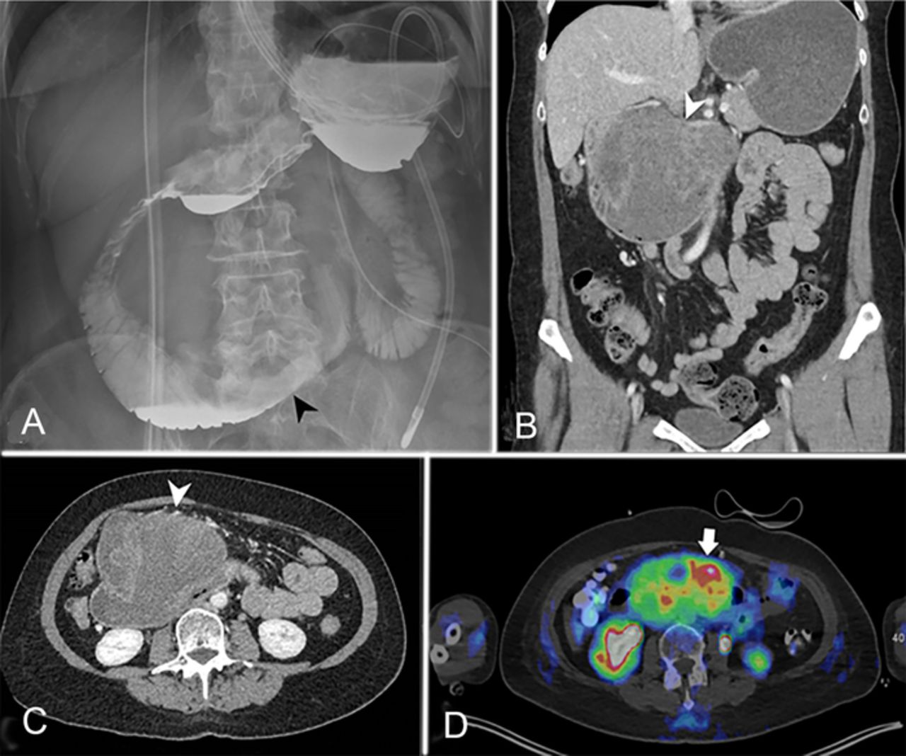
- Figure 1.
(A) Anteroposterior view of the abdomen after administration of oral contrast (Omnipaque, GE Healthcare) demonstrates narrowed third and fourth segments of the duodenum secondary to mass effect (black arrowhead). (B) Coronal and (C) axial computed tomography (CT) images demonstrate a heterogeneous mass arising from the superior and anterior walls of the third segment of the duodenum (white arrowheads). Approximately 90 mL of Visipaque 320 (GE Healthcare) was administered intravenously. (D) Fluorodeoxyglucose (F 18-FDG) positron emission tomography/CT demonstrates the same mass with intense metabolic activity (white arrow). Approximately 7.15 mCi of F 18-FDG was administered intravenously. The patient's blood glucose level just prior to imaging was 90 mg/dL. Time from F 18-FDG injection to scan was 51 minutes.
- Figure 2.
Gross examination demonstrated a 5.2-cm tumor posttreatment (arrowhead) in the right retroperitoneum affixed to the duodenum. The long single suture marks the distal surgical margin (asterisk), and the short single suture marks the proximal surgical margin (black arrow). The double suture on the left represents the tumor margin abutting the superior mesenteric artery (white arrows).
- Figure 3.
Histologic and immunohistochemistry features of the retroperitoneal tumor. (A) Solid and sheet-like configurations of densely packed tumor cells demonstrate indistinct cell borders, scant cytoplasm, fine chromatin, and inconspicuous nucleoli (hematoxylin and eosin, magnification ×100). The boxed region in the upper left corner shows a tumor cell undergoing mitosis (magnification ×400). The tumor is positive for (B) CD99 and negative for (C) vimentin and (D) WT1 (nonnuclear staining) (magnification ×200).


Tables
Study Age/Sex Described Location Unique Imaging Features Unique Pathologic Features Size Adair et al, 200113 21/F Duodenum and jejunum Intussusception of the duodenum N/R 6 × 6 × 4 cm Kie et al, 200311 20/F First and second portion of the duodenum N/R Invasion of the whole layer of duodenum and focal extension to the pancreatic head 6.5 cm Huang et al, 202114 41/M Descending duodenum Enhancing on CT N/R N/R Present case, 2022 66/F Transverse duodenum Heterogenous on CT; intense FDG uptake on PET-CT Multiloculated hemorrhagic mass 10.3 × 8.8 × 12.3 cm CT, computed tomography; F, female; FDG, fluorodeoxyglucose; M, male; N/R, not reported; PET, positron emission tomography.
In this issue


Jump to section
Cited By...
- No citing articles found.