Article Text

Statistics from Altmetric.com
Description
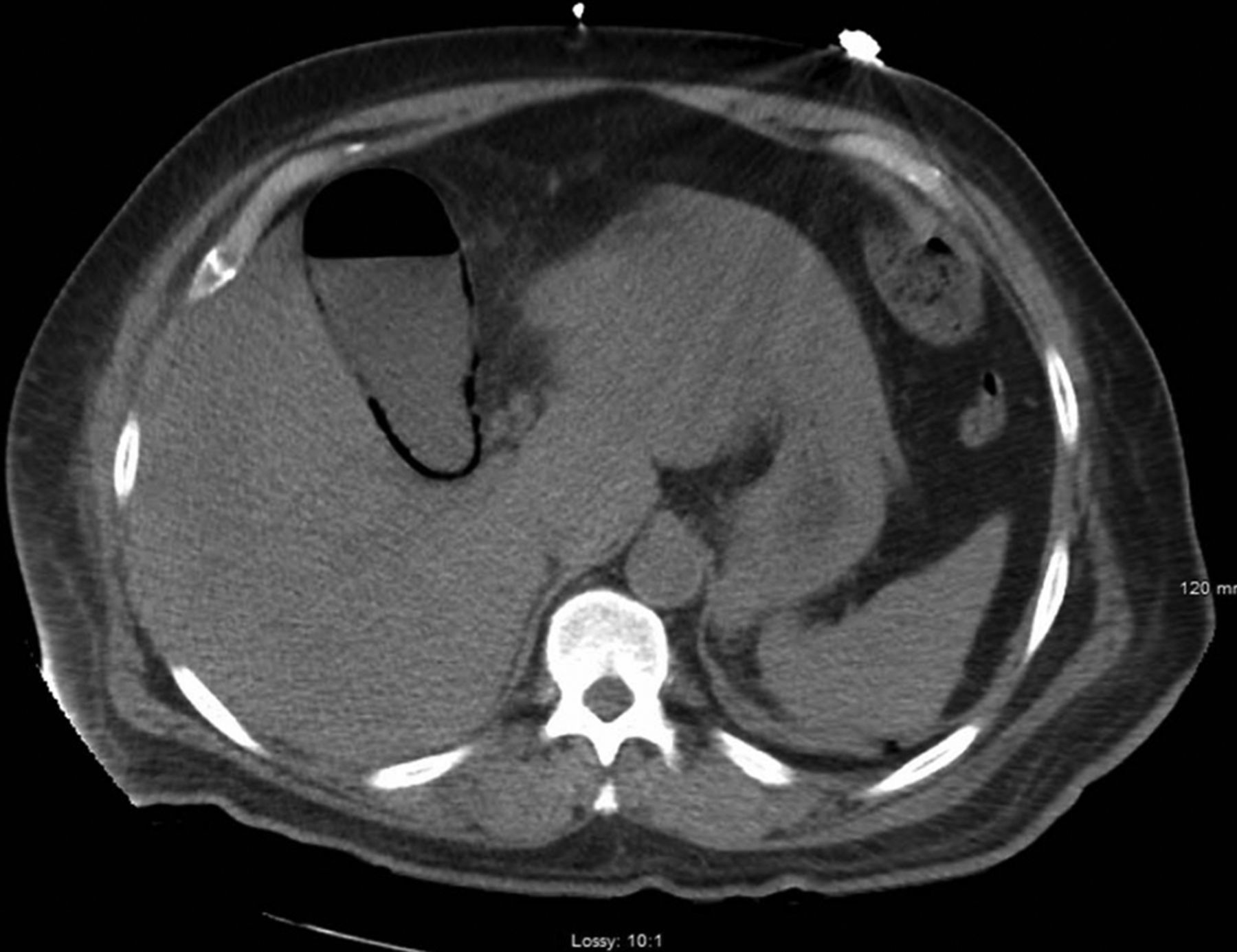
A 62-year-old man with a history of hypertension and poorly controlled type II diabetes mellitus presented to his primary care physician with a 5-day history of chills, decreased urinary output and haematuria. Urinalysis was consistent with urinary tract infection and the patient was prescribed ciprofloxacin. He did not take the antibiotics as instructed and subsequently had to come to Emergency Room due to worsening of his condition as well as new complaints of increased thirst, dark urine and lower back pain. He also admitted that for the previous 3 days he had not been taken insulin due to his illness. On examination, he was an obese man in no distress, fully alert and oriented. The temperature was 97oF, the blood pressure 125/58 mm Hg, the pulse 112 beats/min, the respiratory rate 16 breaths/min and the oxygen saturation 93% on ambient air. Abnormal findings included scleral icterus, mild jaundice, tachycardia, right upper abdominal and bilateral flank tenderness. On initial blood analysis, the patient was found to have leucocytosis, hyperglycaemia, lactate acidosis and hyperbilirubinaemia. His urinalysis results again supported the diagnosis of urinary tract infection. CT scan of the abdomen and pelvis demonstrated bilateral emphysematous pyelonephritis (figure 1), portal venous gas and emphysematous cholecystitis (figure 2). The patient was initially managed with intravenous fluid resuscitation, piperacillin/tazobactam and vancomycin. He was admitted to Medical Intensive Care Unit (MICU) with consultation from general surgery, urology, infectious disease and nephrology. Blood and urine cultures ultimately grew Extended-Spectrum Beta-Lactamase-(ESBL)-producing Escherichia coli and the antibiotics were changed to ertapenem based on sensitivities. He underwent percutaneous cholecystostomy drainage and successful percutaneous drainage of the left kidney avoiding nephrectomy. Both specimens were cultured and grew ESBL E. coli. His renal function deteriorated and he became dependent on haemodialysis. After 10 days, he was deemed stable enough for surgery and underwent cholecystectomy for what was found to be gangrenous cholecystitis. After a prolonged MICU course and hospitalisation, he survived to be discharged to a nursing facility. He eventually recovered his renal function after several months and came off haemodialysis.
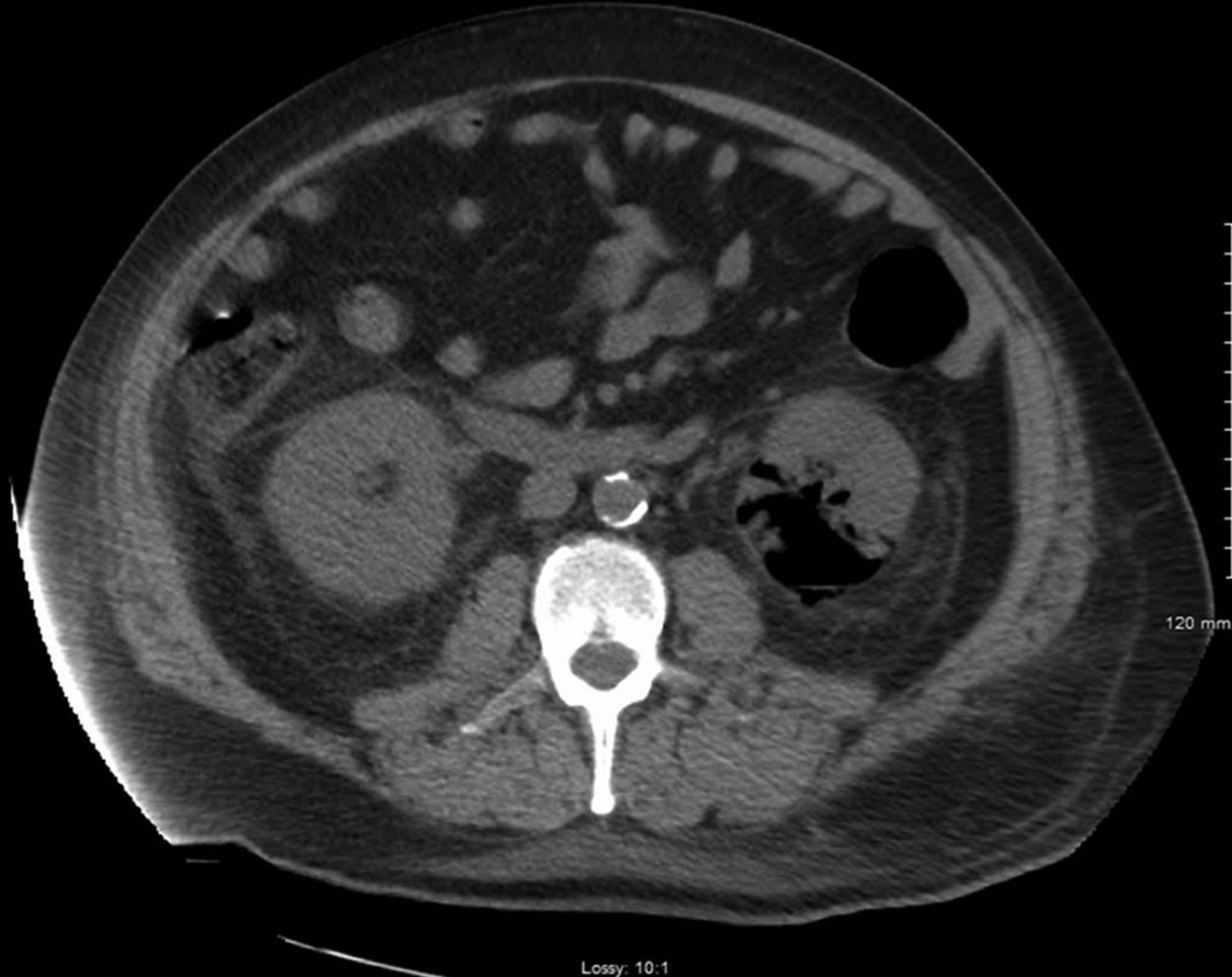
Axial CT image of the abdomen illustrating a large amount of air in the left renal parenchyma with perirenal fat stranding.

Axial CT image of the abdomen demonstrating intramural gas within the gallbladder.
Learning points
Emphysematous pyelonephritis (EP) predominantly affects patients with poorly controlled diabetes mellitus. The imaging modality of choice is CT of the abdomen as it allows a more accurate assessment of the extent of parenchymal gas. Localised disease can be managed with broad spectrum antibiotics and percutaneous nephrostomy or partial nephrectomy, whereas extensive parenchymal involvement with severely impaired renal function necessitates total nephrectomy.
Emphysematous cholecystitis (EC) is necrotising infection of the gallbladder usually seen in diabetic and is associated with high mortality rate of about 15% as compared with only 4% in non-EC. CT scan is the most sensitive and specific imaging modality for identifying gas within gallbladder lumen or wall. Management of EC requires cholecystectomy in conjunction with intravenous fluids and broad-spectrum antibiotics.
Concurrent presentation of EP and EC has scarcely been reported in the literature. The clinical presentation in our case suggests that the infective process initiated as acute cystitis and then progressed to pyelonephritis with ensuing bacteraemia and eventually a secondary cholecystitis. Culture results obtained from the blood, nephrostomy drainage and surgical specimens were all concordant supporting the hypothesis that EP and EC represented evolution of the same septic process.
Footnotes
Contributors CH and EK were equally involved in the concept, literature review and compilation of this case report.
Funding The authors have not declared a specific grant for this research from any funding agency in the public, commercial or not-for-profit sectors.
Competing interests None declared.
Patient consent Obtained.
Provenance and peer review Not commissioned; externally peer reviewed.